Latest Post
Showing posts with label MEDICAL IMAGING. Show all posts

New technology may identify tiny strains in body tissues before injuries occur

Written By Unknown on Friday, January 16, 2015 | 9:13 PM

The top image shows how the new algorithm is able to identify an area (in red) where stress has created a weak spot in a small piece of plastic wrap. The older method (shown in the bottom half of the picture) is unable to pinpoint the place where the plastic wrap is weakening.
Credit: John Boyle, © The Royal Society (used with permission)
Researchers at Washington University in St. Louis have developed algorithms to identify weak spots in tendons, muscles and bones prone to tearing or breaking. The technology, which needs to be refined before it is used in patients, one day may help pinpoint minor strains and tiny injuries in the body's tissues long before bigger problems occur.

The research is available online Aug. 27 in the Journal of the Royal Society Interface, which publishes research at the nexus of the physical and life sciences.

"Tendons are constantly stretching as muscles pull on them, and bones also bend or compress as we carry out everyday activities," said senior investigator Stavros Thomopoulos, PhD, professor of orthopaedic surgery. "Small cracks or tears can result from these loads and lead to major injuries. Understanding how these tears and cracks develop over time therefore is important for diagnosing and tracking injuries."

To that end, Thomopoulos and his colleagues developed a way to visualize and even predict spots where tissues are weakened. To accomplish this, they stretched tissues and tracked what happened as their shapes changed or became distorted.

The paper's first author, John J. Boyle, a graduate student in biomedical engineering, combined mechanical engineering fundamentals with image-analysis techniques to create the algorithms, which were tested in different materials and in animal models.

"If you imagine stretching Silly Putty or a swimming cap with a picture on it, as you pull, the picture becomes distorted," Boyle said. "This allows us to track how the material responds to an external force."

In one of the experiments described in the paper, Boyle sprayed a pattern of dots on plastic wrap, stretched it and tracked the dots.

"As you pull and stretch the plastic wrap, eventually tears begin to emerge," he explained. 

"The new algorithm allowed us to find the places where the tears were beginning to form and to track them as they extended. Older algorithms are not as good at finding and tracking localized strains as the material stretches."

In fact, one of the two new algorithms is 1,000 times more accurate than older methods at quantifying very large stretches near tiny cracks and tears, the research showed. And a second algorithm has the ability to predict where cracks and failures are likely to form.

"This extra accuracy is critical for quantifying large strains," said Guy Genin, PhD, professor of mechanical engineering and co-senior investigator on the study. "Commercial algorithms that estimate strain often are much less sensitive, and they are prone to detecting noise that can arise from the algorithm itself rather than from the material being examined. The new algorithms can distinguish the noise from true regions of large strains."

Thomopoulos, who also is a professor of biomedical engineering and of mechanical engineering, works with Genin to study the shoulder's rotator cuff, a group of tendons and muscles that connect the upper arm to the shoulder blade. They want to learn why some surgeries to repair rotator cuff injuries ultimately fail. Their goal is to increase the odds that the tissue in the shoulder will heal following surgery, and they believe the new algorithms could help them get closer to that goal.

How soon the new algorithms could be used in patients depends on getting better images of the body's tissues. Current imaging techniques, such as MRI and ultrasound, lack the required clarity and resolution.

Genin also explained that although the goal of the current study is to better understand how forces at work on human tissue cause injury and stress, the algorithms also could help engineers identify vulnerable parts of buildings and other structures. Our muscles and bones, he said, are influenced by the same strains that affect those structures.

"Whether it's a bridge or a tendon, it's vital to understand the ways that physical forces cause structures and tissues to deform so that we can identify the onset of failures and eventually predict them," he said.

In the long run, they want to use the algorithms to prevent additional injuries following surgery to repair knees, shoulders and other tissues. They also said it may be possible some day to predict problems before they occur.

The group, which applied for a provisional patent earlier this year, hopes the algorithms will be useful to researchers in the medical and engineering fields.

As a piece of plastic wrap is stretched, the new algorithms identify the location (in red) where it is weakening, which is where the material eventually breaks.

Technique for cardiovascular diagnostics shows promise

Written By Unknown on Thursday, January 15, 2015 | 6:40 PM

Researcher Elira Maksuti is involved in developing and testing a promising method for diagnosing atherosclerosis, or hardening of the arteries. Credit: Staffan Larsson
A new technique developed at Sweden's KTH Royal Institute of Technology shows promise for early diagnosis and treatment of cardiovascular disease.

Hardening of the arteries, or atherosclerosis, is a common disorder that occurs when fat, cholesterol, and other substances build up in the walls of arteries and form hard structures called plaques. The condition can lead to heart attacks and strokes.

To diagnose atherosclerosis, doctors today rely on ultrasonic grayscale images to visually assess vascular function and how very large arteries move. The less mobility, the more developed the case of atherosclerosis.

But it is an indirect measurement. Better diagnoses can be made when the stiffness of the blood vessels can be analysed, explains Elira Maksuti, a researcher at the Department of Medical Imaging Technology at KTH.

"You need a doctor who is an expert and has extensive experience in order to get a good diagnosis," she says.

But by combining the technologies of shear wave elastography and ultrasound, Maksuti and researcher Erik Widmanh, have developed an inexpensive and non-invasive method not only for checking the stiffness of blood vessels, but for analysing the type of plaque present in the artery.

Maksuit says their method not only offers a potentially more effective way to diagnose atherosclerosis, but the ultrasound technology that it relies on is less expensive -- and safer -- than other imaging alternatives, such as magnetic resonance imaging (MRI) or computed tomography (CT).

The technique was tested on artificial blood vessels, or "phantom" vessels, which allowed the researchers to experiment with not only vascular stiffness, but also pressure and flow.

Maksuti says that with the success of tests on these phantoms, the next step is testing the technique with blood vessels from pigs. "These tests also look very promising," she says.

"We see two major future applications before us," she says. "The first is to determine when a patient's blood vessels are becoming rigid, that is, when the atherosclerosis process begins.

"The second application is to be able to diagnose the type of calcification -- or plaque -- present in the blood vessel." Not all plaque is the same: it ranges from hard to soft. If the plaque is soft and has a thin, hard shell, it is more likely to come loose inside the blood vessel.

It's a difficult distinction to determine. But the information is critical in deciding whether to open the artery surgically. "Today it is rather a matter of guessing. A doctor cannot know," she says. And to complicate matters, such operations can also generate strokes.

Source: KTH The Royal Institute of Technology

Powerful imaging for optical point-of-care diagnostics

The new imaging system consists of a handheld probe (on the right), and an ultrasound scanning display system (on the left). It can be easily transported between rooms in a clinic. Credit: Pim van den Berg/ Khalid Daoudi
A new handheld probe developed by a team of university and industry researchers in the Netherlands and France could give doctors powerful new imaging capabilities right in the palms of their hands. The imaging system, which is described in a paper published in The Optical Society's (OSA) open-access journal Optics Express, shrinks a technology that once filled a whole lab bench down to a computer screen and a small probe about the size of a stapler.

The new device combines two imaging modalities: ultrasound and photoacoustics. Ultrasound is a well-established technology that analyzes how sound pulses echo off internal body parts. It is good at revealing anatomical structures and is, perhaps most familiarly, used to image a developing fetus in a mother's womb.

Photoacoustics is a relatively new imaging technique, still making its way toward widespread clinical applications. In photoacoustic imaging, short pulses of light heat up internal tissue. The slight temperature change leads to a change in pressure,
which in turn produces a wave of ultrasound that can be analyzed to reveal information about the body's internal workings. Since this technique ultimately produces ultrasound waves as well, existing technology can be used to analyze and display the images.

The advantage of photoacoustics is that it can reveal important medical information that other imaging techniques cannot, including the presence of molecules like hemoglobin and melanin and the sub-millimeter structure of networks of blood vessels several centimeters beneath the skin. When combined with spectroscopic measurements, photoacoustics can also quantify hemoglobin oxygen saturation within single vessels, providing metabolic information that could be helpful for monitoring tumor progression, for example.

Yet despite these benefits, the cost and size of most photoacoustic systems limit their widespread use, said Khalid Daoudi, a researcher in the Biomedical Photonic Imaging Group at the University of Twente in the Netherlands. Most systems on the market require costly and bulky lasers that make the systems impractical for point-of-care diagnostics. "Our research aimed to break through these hindering factors," Daoudi said.

The project started as collaboration between the University of Twente and three European companies: ESAOTE Europe, a maker of medical diagnostic systems, Quantel, a maker of solid state lasers, and SILIOS Technologies, a maker of optical components.

The team's key innovation, which allowed them to dramatically shrink the system, was the design of an ultra-compact laser based on an efficient and inexpensive laser diode. By stacking multiple diodes to increase the power and carefully designing optical elements to shape the laser beam, the team was able to generate laser pulses with energies higher than had ever been achieved before with diode technology.

Diode lasers can also provide many laser pulses per second, which in turn allows real time imaging, another advantage of the new system, Daoudi noted.

The researchers tested the imaging performance of the system in different types of phantoms -- materials designed to mimic a tissue's optical properties -- and in a healthy human finger joint.

The new compact probe and imaging system can be easily transported between rooms in a clinical setting, an attractive feature for future commercialization, the researchers said.

The team is currently working with a European consortium of industrial and academic partners to take the next steps from the research to the commercialization phase. The current system operates at a single wavelength in the near infrared, but the team has plans to expand the design to multi-wavelength imaging.

"Some applications targeted are rheumatoid arthritis in finger joints, oncology, cardiovascular disease and burn wounds," Daoudi said.

Source: The Optical Society

Ultrasound guides tongue to pronounce 'R' sounds

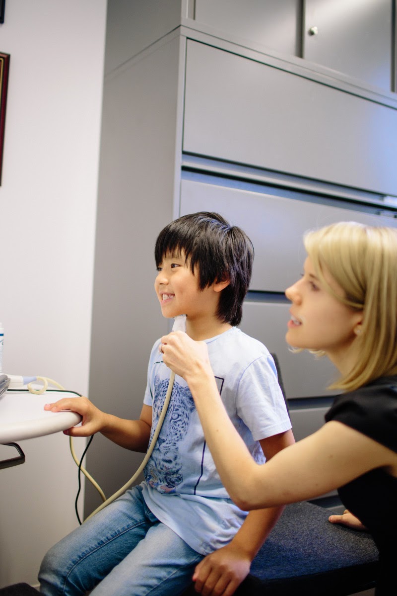
Using ultrasound technology to visualize the tongue's shape and movement can help children with difficulty pronouncing "r" sounds, according to research led by NYU Steinhardt assistant professor Tara McAllister Byun. Credit: Ramsay de Give / NYU Steinhardt
Using ultrasound technology to visualize the tongue's shape and movement can help children with difficulty pronouncing "r" sounds, according to a small study by NYU's Steinhardt School of Culture, Education, and Human Development and Montclair State University.

The ultrasound intervention was effective when individuals were allowed to make different shapes with their tongue in order to produce the "r" sound, rather than being instructed to make a specific shape. The findings appear online in the Journal of Speech, Language, and Hearing Research.

The "r" sound is one of the most frequent speech errors, and can be challenging to correct. For other sounds -- such as "t" or "p" -- speech pathologists can give clear verbal, visual or tactile cues to help children understand how the sound is created. "R" is difficult to show or describe in an easy-to-understand fashion.

In addition, most speech sounds are produced in the same way, but with "r," normal speakers use widely different tongue shapes to create the sound. The two primary strategies to create the "r" sound include a retroflex tongue shape, where the tongue tip is pointed up, and the bunched tongue shape, where the tongue tip is pointed down and body of tongue bunches up toward the top of the mouth.

Up to 10 percent of children have speech sound disorders, according to the National Institutes of Health. Some children respond well to conventional forms of speech therapy, but others have errors that persist despite their therapists' best efforts. A growing body of evidence suggests that treatment incorporating visual biofeedback, which uses various technologies to create a dynamic visual representation of speech, could fill this need.

"The idea that you could get around the challenges with 'r' sounds by showing children their tongues as they are talking is really appealing to clinicians," says Tara McAllister Byun, an assistant professor in NYU Steinhardt's Department of Communicative Sciences and Disorders and the study's lead author. "That's what ultrasound technology lets us do."

Linguists have used ultrasound in the past to study basic functions of speech, and in recent years, speech pathologists have begun exploring using ultrasound to treat children with speech errors. An ultrasound probe -- similar to ones used in cardiac and tissue imaging -- is held under the chin, and sound waves capture real-time images of the tongue. The images provide both the child and speech pathologist with information about the tongue's position and shape.

Using the ultrasound images as a guide, children learn how to manipulate their tongues, and speech pathologists advise them on how to make adjustments to better achieve different sounds.

Several case studies and small studies suggest that ultrasound biofeedback can successfully correct "r" speech errors. Byun and her colleagues set out to gather systematic evidence on the effectiveness of the treatment, studying eight children with difficulty pronouncing "r" sounds. Seven of the eight had previous speech therapy that was unsuccessful.

Four children participated in the initial eight-week study. They were taught to make a bunched tongue shape, guided by ultrasound, in an effort to better pronounce "r." The researchers saw only small improvements among the four participants.

However, while trying to create a bunched tongue, one child stumbled upon a retroflex tongue shape and was able to improve her "r" sound. As a result of her success, the researchers altered their study design to allow participants to choose their own tongue shape, with individualized guidance from speech language pathologists.

A different four children participated in the second study over an eight-week period. Using ultrasound to visualize their tongues, all four participants in the second study showed significant improvement in their "r" sounds.

"Our second study offers evidence that when flexibility is given to choose a tongue shape, rather than a one-size-fits all approach, ultrasound biofeedback treatment can be a highly effective intervention for children with trouble pronouncing 'r' sounds," Byun says.

The researchers noted that the two studies were not a controlled comparison, thus additional systematic research is needed before drawing strong conclusions about the importance of individualized tongue shapes.

New biometric watches use light to non-invasively monitor glucose, dehydration, pulse

Written By Unknown on Wednesday, January 14, 2015 | 5:42 AM

This schematic diagram shows how the new system can be used to measure a person's glucose levels noninvasively. Credit: Biomedical Optics Express
Monitoring a patient's vital signs and other physiological parameters is a standard part of medical care, but, increasingly, health and fitness-minded individuals are looking for ways to easily keep their own tabs on these measurements. Enter the biometric watch.

In a pair of papers published in The Optical Society's (OSA) open-access journal Biomedical Optics Express, groups of researchers from the Netherlands and Israel describe two new wearable devices that use changing patterns of scattered light to monitor biometrics: one tracks glucose concentration and dehydration levels, and the other monitors pulse.

The glucose sensor is the first wearable device that can measure glucose concentration directly but noninvasively, the authors say.
And while other wearable devices have been made to monitor pulse, the authors claim their new design would be less sensitive to errors when the wearer is in motion, for example while walking or playing sports

Both of the watches described in the two papers make use of the so-called "speckle" effect, the grainy interference patterns that are produced on images when laser light reflects from an uneven surface or scatters from an opaque material. When the material that is scattering the light is moving -- say, in the case of blood flowing through the circulatory system -- "the speckle pattern changes with changes in the flow," explained biomedical engineer Mahsa Nemati, a graduate student in the Optics Research Group at the Delft University of Technology in the Netherlands and the lead author of the Biomedical Optics Express paper on monitoring pulse. Those light variations are a valuable source of information, she says.

The 'Holy Grail' of Diagnostics

In the first paper, bioengineer Zeev Zalevsky of Israel's Bar-Ilan University and his colleagues describe a new wearable biometric system that uses the speckle effect to directly monitor the glucose concentration in the bloodstream, as well as the wearer's relative hydration level.

"Glucose is the holy grail of the world of biomedical diagnostics, and dehydration is a very useful parameter in the field of wellness, which is one of our main commercial aims," Zalevsky said.

The watch-like device consists of a laser to generate a wavefront of light that illuminates a patch of skin on the wrist near an artery, and a camera that measures changes over time in the light that is backscattered off the skin. Unlike other chemicals present in the blood, glucose exhibits a so-called Faraday effect. This means that in the presence of an external magnetic field (generated by a magnet attached to the device) the glucose molecule alters the polarization of the wavefront and thus influences the resulting speckle patterns. 
Analyzing these changing patterns provides a direct measurement of the glucose concentration. Because one of the main signs of mild to moderate dehydration is muscle weakness, which will alter the strength of the signals, the same device can also be used to indicate the relative dehydration level of the user as it changes over time.

Zalevsky and his colleagues are now working to reduce the margin of error in the device's readings. "Around 96 percent of our in vivo measurements were within a range of 15 percent deviation from the readout of a medical reference glucometer device," Zalevsky noted. "The main factor for errors now is the stability of our device on the wrist of the user. We are currently investing efforts in deriving proper calibration and motion cancellation procedures that will allow us to reduce this sensitivity."

Zalevsky says this is the first step toward non-invasive, continuous in vivo measurement of glucose that is based on sensing an effect that is directly related to glucose concentration. The team expects a commercial version of the device to reach the market within two to three years.

Pulse Tracker

In the second Biomedical Optics Express paper, Nemati and her colleagues at Delft and at Phillips Research developed a method that could be used to monitor pulse non-invasively with a sensor that isn't thrown off by the wearer's movement.

Using simulated heart beats generated in milk and measurements performed on the finger of a volunteer, they found that speckle changes can be used to accurately measure flow pulsations -- that is, the heart rate -- even when the light source used to create the speckle pattern is also moving, as would be the case with a wearable biometric sensor. The researchers found that just a couple of pixels from the image were sufficient to extract the pulse rate.

"This paper shows for the first time that a speckle pattern generated from a flowing liquid can give us the pulsation properties of the flow in spite of motion-induced artifacts," Nemati said. "Sophisticated optics is not necessary to implement this, so the costs for devices can be kept low. Another advantage is that the devices can be non-contact or far from the sample," she added.

The team is currently working with companies to integrate their motion-friendly pulse-monitoring technique into existing sensors, for potential use clinically as well as in sports, Nemati said.

Source: The Optical Society
 
Support : Creating Website | Johny Template | Mas Template
Copyright © 2011. The planet wall - All Rights Reserved
Template Created by Easy Blogging Published by Mas Template
Proudly powered by Blogger