Latest Post
Showing posts with label ULTRASOUND NEWS. Show all posts

Technique for cardiovascular diagnostics shows promise

Written By Unknown on Thursday, January 15, 2015 | 6:40 PM

Researcher Elira Maksuti is involved in developing and testing a promising method for diagnosing atherosclerosis, or hardening of the arteries. Credit: Staffan Larsson
A new technique developed at Sweden's KTH Royal Institute of Technology shows promise for early diagnosis and treatment of cardiovascular disease.

Hardening of the arteries, or atherosclerosis, is a common disorder that occurs when fat, cholesterol, and other substances build up in the walls of arteries and form hard structures called plaques. The condition can lead to heart attacks and strokes.

To diagnose atherosclerosis, doctors today rely on ultrasonic grayscale images to visually assess vascular function and how very large arteries move. The less mobility, the more developed the case of atherosclerosis.

But it is an indirect measurement. Better diagnoses can be made when the stiffness of the blood vessels can be analysed, explains Elira Maksuti, a researcher at the Department of Medical Imaging Technology at KTH.

"You need a doctor who is an expert and has extensive experience in order to get a good diagnosis," she says.

But by combining the technologies of shear wave elastography and ultrasound, Maksuti and researcher Erik Widmanh, have developed an inexpensive and non-invasive method not only for checking the stiffness of blood vessels, but for analysing the type of plaque present in the artery.

Maksuit says their method not only offers a potentially more effective way to diagnose atherosclerosis, but the ultrasound technology that it relies on is less expensive -- and safer -- than other imaging alternatives, such as magnetic resonance imaging (MRI) or computed tomography (CT).

The technique was tested on artificial blood vessels, or "phantom" vessels, which allowed the researchers to experiment with not only vascular stiffness, but also pressure and flow.

Maksuti says that with the success of tests on these phantoms, the next step is testing the technique with blood vessels from pigs. "These tests also look very promising," she says.

"We see two major future applications before us," she says. "The first is to determine when a patient's blood vessels are becoming rigid, that is, when the atherosclerosis process begins.

"The second application is to be able to diagnose the type of calcification -- or plaque -- present in the blood vessel." Not all plaque is the same: it ranges from hard to soft. If the plaque is soft and has a thin, hard shell, it is more likely to come loose inside the blood vessel.

It's a difficult distinction to determine. But the information is critical in deciding whether to open the artery surgically. "Today it is rather a matter of guessing. A doctor cannot know," she says. And to complicate matters, such operations can also generate strokes.

Source: KTH The Royal Institute of Technology

Early results indicate potential for focused ultrasound to treat OCD

Dr. Chang (in sweater) and treatment team with the first patient in the OCD study. Credit: Image courtesy of Focused Ultrasound Foundation
A recently published report in the Journal of Molecular Psychiatry supports the potential of focused ultrasound to treat certain patients with obsessive-compulsive disorder (OCD).

In a study of feasibility, safety and preliminary efficacy, four patients with disabling OCD unresponsive to medical therapy were treated by ablating small targets deep in the brain (anterior internal capsule) using the InSightec ExAblate Neuro MR-guided focused ultrasound system. This proof-of-concept study is being conducted with 12 patients by Jin Woo Chang, MD, PhD, at the Yonsei University Medical Center in Seoul, Korea. The results of the first four patients with six months follow-up were published.

All four patients had the targeted areas of the brain successfully ablated. The procedures were accomplished without complications or side effects and resulted in gradual improvement in obsessive-compulsive thoughts and behaviors as well as showing the nearly immediate and sustained improvement in depression and anxiety over six months.

"There is a need for non-invasive treatment options for patients with OCD that cannot be managed through medication," says Dr. Chang. "Using focused ultrasound, we were able to reduce the symptoms for these patients and help them get some of their life back without the risks or complications of the more invasive surgical approaches that are currently available."

"If these initial results are confirmed in the remaining eight patients in this study as well as in a larger pivotal trial of safety and efficacy, focused ultrasound could emerge as an alternative to surgery for improving quality of life in a cost-effective manner for patients with OCD," said Foundation Chairman Neal F. Kassell, MD. "This could also serve as the predicate for non-invasive therapy for other psychiatric disorders."

Dr. Chang plans to start a study using focused ultrasound to treat depression in 2015.

Current Treatment for OCD

Obsessive-compulsive disorder (OCD) is a psychiatric disorder characterized by recurring anxiety-provoking thoughts (obsessions) that are alleviated only by ritualistic actions (compulsions) such as hand washing. OCD symptoms can be overwhelming and result in severe impairment and dysfunction. Patients also have a high risk for depression, with two-thirds of OCD patients developing major depression.

Although a large portion of OCD patients improve with drug therapy, some patients have debilitating symptoms that are resistant to treatment. For these patients, psychosurgery can be performed to destroy (ablate) a targeted region of the brain (anterior internal capsule) associated with the disorder. The currently available ablative approaches are invasive or involve radiation:

• Radiofrequency ablation requires drilling into the skull and inserting a probe through 
   the brain.

• Stereotactic radiosurgery takes months to achieve an effect and may be associated with
   side effects of radiation.

• Deep brain stimulation has been used as a reversible and adjustable alternative to ablation, but it involves the risks and complications of surgery to implant the system and additional operations for battery changes.

Source: Focused Ultrasound Foundation

Ultrasounds dance the 'moonwalk' in new metamaterial

Silicone beads embedded in a water-based gel (photograph is ~2 cm across). Credit: © CRPP
Metamaterials have extraordinary properties when it comes to diverting and controlling waves, especially sound and light: for instance, they can make an object invisible, or increase the resolving power of a lens. Now, researchers at the Centre de Recherche Paul Pascal (CNRS) and the Institut de Mรฉcanique et d'Ingรฉnierie de Bordeaux (CNRS/Universitรฉ de Bordeaux/Bordeaux INP/Arts et Mรฉtiers ParisTech) have developed the first three-dimensional metamaterials by combining physico-chemical formulation and microfluidics technology. This is a new generation of soft metamaterials that are easier to shape. In their experiment, the researchers got ultrasonic oscillations to move backwards while the energy carried by the wave moved forwards. Their work opens up new prospects, especially for high-resolution imaging (ultrasonography). It is published on 15 December 2014 in the journal Nature Materials.

Since the 2000s, the international scientific community has seen interest in metamaterials and their extraordinary properties grow exponentially. A metamaterial is a medium in which the phase velocity of light or sound waves can be negative (the material is said to have a negative refractive index).. In such a medium, the phase of the wave (the successive oscillations) and the energy carried by this same wave move in opposite directions. This property is not found in any natural homogeneous medium.

To obtain a metamaterial, it is necessary to make a heterogeneous medium that contains a large number of inclusions (known as microresonators). The usual way is to use micromechanical methods (etching, deposition, etc) to machine solid supports that will have the properties of metamaterials in one or two dimensions. However, this method cannot be used to work with soft matter at the micrometer scales required for ultrasounds, and the materials obtained remain limited to one or two dimensions.

In this study, the researchers developed a new type of metamaterial, in the fluid phase, formed of porous silicone microbeads embedded in a water-based gel. This metafluid is the first three-dimensional metamaterial to work at ultrasonic frequencies. In addition, due to its fluid nature, it can be made using physico-chemical processes and microfluidics technologies, which are much easier to implement than micromechanical methods.

One of the properties of porous media is that sound travels through them at very low speed (a few tens of meters per second) compared to water (1500 meters per second). Due to this sharp contrast, the whole suspension has the properties of a metamaterial provided the bead concentration is sufficient: when the researchers studied the propagation of ultrasonic waves through this medium, they directly measured a negative refractive index. Within such a metafluid, the energy carried by the wave travels from the emitter to the receiver, as expected, whereas the oscillations appear to move backwards in the opposite direction, rather like a dancer doing the 'moonwalk'.

These results open the way to numerous applications ranging from high-resolution ultrasound imaging to sound insulation and stealth in underwater acoustics. In addition, the soft-matter physico-chemical techniques used to make this metamaterial makes it possible to produce fluid or flexible materials with adaptable shapes, potentially at the industrial scale.

High-intensity sound waves may aid regenerative medicine

This is a cross section through a histotripsy lesion created in bovine liver tissue with the liquified cellular contents washed out revealing the remaining extracellular matrix. The scale bar represents 5mm. Credit: T.Khoklova/UW
Researchers at the University of Washington have developed a way to use sound to create cellular scaffolding for tissue engineering, a unique approach that could help overcome one of regenerative medicine's significant obstacles. The researchers will present their technique at the 168th meeting of the Acoustical Society of America (ASA), held October 27-31, 2014, at the Indianapolis Marriott Downtown Hotel.

The development of the new technique started with somewhat of a serendipitous discovery. The University of Washington team had been studying boiling histotripsy -- a technique that uses millisecond-long bursts of high-intensity ultrasound waves to break apart tissue -- as a method to eliminate cancerous tumors by liquefying them with ultrasound waves. After the sound waves destroy the tumors, the body should eliminate them as cellular waste. When the researchers examined these 'decellularized' tissues, however, they were surprised by what the boiling left intact.

"In some of our experiments, we discovered that some of the stromal tissue and vasculature was being left behind," said Yak-Nam Wang, a senior engineer at the University of Washington's Applied Physics Laboratory. "So we had the idea about using this to decellularize tissues for tissue engineering and regenerative medicine."

The structure that remains after decellularizing tissues is known as the extracellular matrix, a fibrous network that provides a scaffold for cells to grow upon. Most other methods for decellularizing tissues and organs involve chemical and enzymatic treatments that can cause damage to the tissues and fibers and takes multiple days. Histrostipsy, on the other hand, offers the possibility of fast decellularization of tissue with minimal damage to the matrix.

"In tissue engineering, one of the holy grails is to develop biomimetic structures so that you can replace tissues with native tissue," Wang said. Stripping away cells from already developed tissue could provide a good candidate for these structures, since the extracellular matrix already acts as the cellular framework for tissue systems, Wang said.

Due to its bare composition, the matrix also induces only a relatively weak immune response from the host. The matrix could then theoretically be fed with stem cells or cells from the same person to effectively re-grow an organ.

"The other thought is that maybe you could just implant the extracellular matrix and then the body itself would self-seed the tissues, if it's just a small patch of tissue that you're replacing," Wang said. "You won't have any immune issues, and because you have this biomimetic scaffold that's closer to the native tissue, healing would be better, and the body would recognize it as normal tissue."

Wang is currently investigating decellularization of kidney and liver tissue from large animals. Future work involves increasing the size of the decellularized tissues and assessing their in-vivo regenerative efficacy.

Powerful imaging for optical point-of-care diagnostics

The new imaging system consists of a handheld probe (on the right), and an ultrasound scanning display system (on the left). It can be easily transported between rooms in a clinic. Credit: Pim van den Berg/ Khalid Daoudi
A new handheld probe developed by a team of university and industry researchers in the Netherlands and France could give doctors powerful new imaging capabilities right in the palms of their hands. The imaging system, which is described in a paper published in The Optical Society's (OSA) open-access journal Optics Express, shrinks a technology that once filled a whole lab bench down to a computer screen and a small probe about the size of a stapler.

The new device combines two imaging modalities: ultrasound and photoacoustics. Ultrasound is a well-established technology that analyzes how sound pulses echo off internal body parts. It is good at revealing anatomical structures and is, perhaps most familiarly, used to image a developing fetus in a mother's womb.

Photoacoustics is a relatively new imaging technique, still making its way toward widespread clinical applications. In photoacoustic imaging, short pulses of light heat up internal tissue. The slight temperature change leads to a change in pressure,
which in turn produces a wave of ultrasound that can be analyzed to reveal information about the body's internal workings. Since this technique ultimately produces ultrasound waves as well, existing technology can be used to analyze and display the images.

The advantage of photoacoustics is that it can reveal important medical information that other imaging techniques cannot, including the presence of molecules like hemoglobin and melanin and the sub-millimeter structure of networks of blood vessels several centimeters beneath the skin. When combined with spectroscopic measurements, photoacoustics can also quantify hemoglobin oxygen saturation within single vessels, providing metabolic information that could be helpful for monitoring tumor progression, for example.

Yet despite these benefits, the cost and size of most photoacoustic systems limit their widespread use, said Khalid Daoudi, a researcher in the Biomedical Photonic Imaging Group at the University of Twente in the Netherlands. Most systems on the market require costly and bulky lasers that make the systems impractical for point-of-care diagnostics. "Our research aimed to break through these hindering factors," Daoudi said.

The project started as collaboration between the University of Twente and three European companies: ESAOTE Europe, a maker of medical diagnostic systems, Quantel, a maker of solid state lasers, and SILIOS Technologies, a maker of optical components.

The team's key innovation, which allowed them to dramatically shrink the system, was the design of an ultra-compact laser based on an efficient and inexpensive laser diode. By stacking multiple diodes to increase the power and carefully designing optical elements to shape the laser beam, the team was able to generate laser pulses with energies higher than had ever been achieved before with diode technology.

Diode lasers can also provide many laser pulses per second, which in turn allows real time imaging, another advantage of the new system, Daoudi noted.

The researchers tested the imaging performance of the system in different types of phantoms -- materials designed to mimic a tissue's optical properties -- and in a healthy human finger joint.

The new compact probe and imaging system can be easily transported between rooms in a clinical setting, an attractive feature for future commercialization, the researchers said.

The team is currently working with a European consortium of industrial and academic partners to take the next steps from the research to the commercialization phase. The current system operates at a single wavelength in the near infrared, but the team has plans to expand the design to multi-wavelength imaging.

"Some applications targeted are rheumatoid arthritis in finger joints, oncology, cardiovascular disease and burn wounds," Daoudi said.

Source: The Optical Society

Ultrasound guides tongue to pronounce 'R' sounds

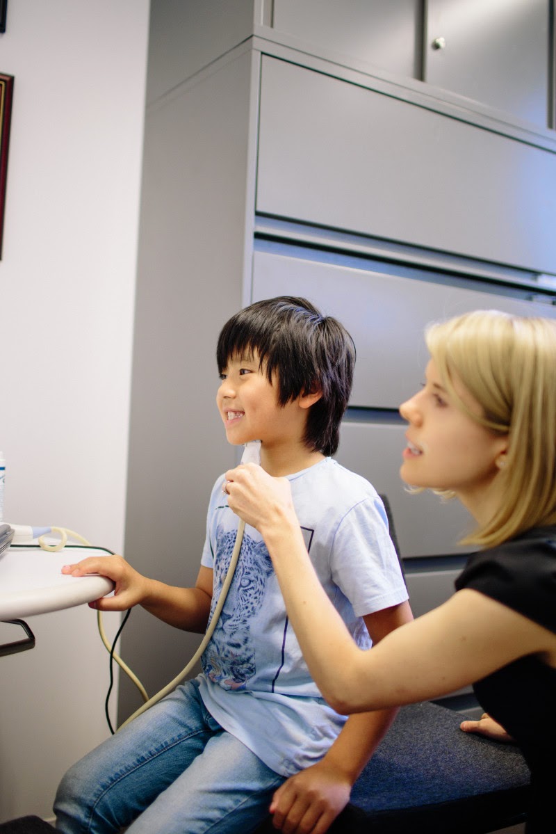
Using ultrasound technology to visualize the tongue's shape and movement can help children with difficulty pronouncing "r" sounds, according to research led by NYU Steinhardt assistant professor Tara McAllister Byun. Credit: Ramsay de Give / NYU Steinhardt
Using ultrasound technology to visualize the tongue's shape and movement can help children with difficulty pronouncing "r" sounds, according to a small study by NYU's Steinhardt School of Culture, Education, and Human Development and Montclair State University.

The ultrasound intervention was effective when individuals were allowed to make different shapes with their tongue in order to produce the "r" sound, rather than being instructed to make a specific shape. The findings appear online in the Journal of Speech, Language, and Hearing Research.

The "r" sound is one of the most frequent speech errors, and can be challenging to correct. For other sounds -- such as "t" or "p" -- speech pathologists can give clear verbal, visual or tactile cues to help children understand how the sound is created. "R" is difficult to show or describe in an easy-to-understand fashion.

In addition, most speech sounds are produced in the same way, but with "r," normal speakers use widely different tongue shapes to create the sound. The two primary strategies to create the "r" sound include a retroflex tongue shape, where the tongue tip is pointed up, and the bunched tongue shape, where the tongue tip is pointed down and body of tongue bunches up toward the top of the mouth.

Up to 10 percent of children have speech sound disorders, according to the National Institutes of Health. Some children respond well to conventional forms of speech therapy, but others have errors that persist despite their therapists' best efforts. A growing body of evidence suggests that treatment incorporating visual biofeedback, which uses various technologies to create a dynamic visual representation of speech, could fill this need.

"The idea that you could get around the challenges with 'r' sounds by showing children their tongues as they are talking is really appealing to clinicians," says Tara McAllister Byun, an assistant professor in NYU Steinhardt's Department of Communicative Sciences and Disorders and the study's lead author. "That's what ultrasound technology lets us do."

Linguists have used ultrasound in the past to study basic functions of speech, and in recent years, speech pathologists have begun exploring using ultrasound to treat children with speech errors. An ultrasound probe -- similar to ones used in cardiac and tissue imaging -- is held under the chin, and sound waves capture real-time images of the tongue. The images provide both the child and speech pathologist with information about the tongue's position and shape.

Using the ultrasound images as a guide, children learn how to manipulate their tongues, and speech pathologists advise them on how to make adjustments to better achieve different sounds.

Several case studies and small studies suggest that ultrasound biofeedback can successfully correct "r" speech errors. Byun and her colleagues set out to gather systematic evidence on the effectiveness of the treatment, studying eight children with difficulty pronouncing "r" sounds. Seven of the eight had previous speech therapy that was unsuccessful.

Four children participated in the initial eight-week study. They were taught to make a bunched tongue shape, guided by ultrasound, in an effort to better pronounce "r." The researchers saw only small improvements among the four participants.

However, while trying to create a bunched tongue, one child stumbled upon a retroflex tongue shape and was able to improve her "r" sound. As a result of her success, the researchers altered their study design to allow participants to choose their own tongue shape, with individualized guidance from speech language pathologists.

A different four children participated in the second study over an eight-week period. Using ultrasound to visualize their tongues, all four participants in the second study showed significant improvement in their "r" sounds.

"Our second study offers evidence that when flexibility is given to choose a tongue shape, rather than a one-size-fits all approach, ultrasound biofeedback treatment can be a highly effective intervention for children with trouble pronouncing 'r' sounds," Byun says.

The researchers noted that the two studies were not a controlled comparison, thus additional systematic research is needed before drawing strong conclusions about the importance of individualized tongue shapes.

 
Support : Creating Website | Johny Template | Mas Template
Copyright © 2011. The planet wall - All Rights Reserved
Template Created by Easy Blogging Published by Mas Template
Proudly powered by Blogger