Latest Post
Showing posts with label SPORTS MEDICINE. Show all posts

How to sell the drugs of the future

Written By Unknown on Thursday, January 15, 2015 | 11:04 PM

Drugs
Credit: Getty Images
Only a decade ago, basing medical treatment on your DNA seemed like science fiction. Not any more. Thanks in part to the sequencing of the human genome, personalized medicine (PM), a specific course of treatment developed for the individual patient, is now science fact.

PM has already shown its effectiveness in the treatment of cancer, and medical professionals are eager to expand it to treat other chronic diseases. But first patients need to understand how PM can work for them.

Will they buy into it? "Yes -- but only if patients are armed with knowledge about their own disease and understand the relative advantages of PM," says Concordia University marketing professor Lea Prevel Katsanis, the co-author of a new study on the subject, published in the International Journal of Pharmaceutical and Healthcare Marketing. She adds that if patients are going to accept PM, doctor-patient communication is vital.

For the study, Katsanis and her co-author, Anja Hitz, a former John Molson School of Business MBA student and current head of medical compliance and prevention at the Military Hospital in Hamburg, Germany, polled 307 consumers through an online survey. 
They found that knowledge and the relative advantages of PM have the most significant influence on patient acceptance of PM.

"The more a patient knows about how she is being treated, the more likely she is to accept that treatment," says Katsanis. "So it's important to educate consumers on potential benefits and risks associated with PM."

Indeed, patient understanding is a key factor in getting healthcare professionals, governments and insurance companies to adopt and pay for PM, particularly when these targeted treatments are often more costly than traditional medical methods.

With PM, the same drug isn't given to millions of people. It's a targeted treatment regime. While that reduced patient pool means an increased cost, there can be long-term benefits. Increased efficiency and prevention may result in fewer drugs being prescribed. And PM may also result in the reduction of secondary costs as a result of overdosing, incorrect prescriptions and adverse drug reactions.

"If PM can be successfully integrated into the healthcare system at a reasonable cost, it represent a significant improvement in the treatment of chronic disease," says Katsanis.

But she warns that marketers need to proceed with caution: "The promotion of personalized medications will increasingly focus on the healthy patient with a genetic disposition for a particular illness. While this might lead to new and potentially greater opportunities for marketers, it might also result in the targeting of healthy patients who don't actually need treatment for an active disease. Ultimately, this could increase healthcare costs and cause unnecessary patient treatment."

Source: Concordia University

High-intensity sound waves may aid regenerative medicine

This is a cross section through a histotripsy lesion created in bovine liver tissue with the liquified cellular contents washed out revealing the remaining extracellular matrix. The scale bar represents 5mm. Credit: T.Khoklova/UW
Researchers at the University of Washington have developed a way to use sound to create cellular scaffolding for tissue engineering, a unique approach that could help overcome one of regenerative medicine's significant obstacles. The researchers will present their technique at the 168th meeting of the Acoustical Society of America (ASA), held October 27-31, 2014, at the Indianapolis Marriott Downtown Hotel.

The development of the new technique started with somewhat of a serendipitous discovery. The University of Washington team had been studying boiling histotripsy -- a technique that uses millisecond-long bursts of high-intensity ultrasound waves to break apart tissue -- as a method to eliminate cancerous tumors by liquefying them with ultrasound waves. After the sound waves destroy the tumors, the body should eliminate them as cellular waste. When the researchers examined these 'decellularized' tissues, however, they were surprised by what the boiling left intact.

"In some of our experiments, we discovered that some of the stromal tissue and vasculature was being left behind," said Yak-Nam Wang, a senior engineer at the University of Washington's Applied Physics Laboratory. "So we had the idea about using this to decellularize tissues for tissue engineering and regenerative medicine."

The structure that remains after decellularizing tissues is known as the extracellular matrix, a fibrous network that provides a scaffold for cells to grow upon. Most other methods for decellularizing tissues and organs involve chemical and enzymatic treatments that can cause damage to the tissues and fibers and takes multiple days. Histrostipsy, on the other hand, offers the possibility of fast decellularization of tissue with minimal damage to the matrix.

"In tissue engineering, one of the holy grails is to develop biomimetic structures so that you can replace tissues with native tissue," Wang said. Stripping away cells from already developed tissue could provide a good candidate for these structures, since the extracellular matrix already acts as the cellular framework for tissue systems, Wang said.

Due to its bare composition, the matrix also induces only a relatively weak immune response from the host. The matrix could then theoretically be fed with stem cells or cells from the same person to effectively re-grow an organ.

"The other thought is that maybe you could just implant the extracellular matrix and then the body itself would self-seed the tissues, if it's just a small patch of tissue that you're replacing," Wang said. "You won't have any immune issues, and because you have this biomimetic scaffold that's closer to the native tissue, healing would be better, and the body would recognize it as normal tissue."

Wang is currently investigating decellularization of kidney and liver tissue from large animals. Future work involves increasing the size of the decellularized tissues and assessing their in-vivo regenerative efficacy.

Football players found to have brain damage from mild 'unreported' concussions

Written By Unknown on Wednesday, January 14, 2015 | 6:20 PM

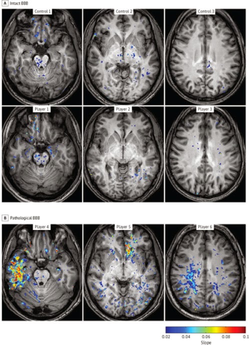
The images from the Ben-Gurion University of the Negev JAMA Neurology study represent Blood-Brain Barrier (BBB) Permeability in Football Players (A) vs. a control group (B). The players in the pathological-BBB group (B) presented focal BBB lesions in different cortical regions including the temporal (player 4), frontal (player 5), and parietal (player 6) lobes. Both gray and white matter were involved. Credit: Image courtesy of American Associates, Ben-Gurion University of the Negev
A new, enhanced MRI diagnostic approach was, for the first time, able to identify significant damage to the blood-brain barrier (BBB) of professional football players following "unreported" trauma or mild concussions. Published in the current issue of JAMA Neurology, this study could improve decision making on when an athlete should "return to play."

According to Prof. Alon Friedman, from the Ben-Gurion University Brain Imaging Research Center and discoverer of the new diagnostic, "until now, there wasn't a diagnostic capability to identify mild brain injury early after the trauma. In the NFL, other professional sports and especially school sports, concern has grown about the long-term neuropsychiatric consequences of repeated mild Traumatic Brain Injury (mTBI) and specifically sports-related concussive and sub-concussive head impacts."

The paper, published by researchers at Ben-Gurion University of the Negev (BGU) and Soroka University Medical Center, describes a new diagnostic approach using Magnetic Resonance Imaging (MRI) for detection and localization of vascular pathology and blood-brain barrier breakdown in football players.

The images from the Ben-Gurion University of the Negev JAMA Neurology study represent Blood-Brain Barrier (BBB) Permeability in Football Players (A) vs. a control group (B). The players in the pathological-BBB group (B) presented focal BBB lesions in different cortical regions including the temporal (player 4), frontal (player 5), and parietal (player 6) lobes. Both gray and white matter were involved.

"The goal of our study was to use our new method to visualize the extent and location of BBB dysfunction in football players using Dynamic Contrast-Enhanced Magnetic Resonance Imaging (DCE-MRI) on a Phillips 3-T Ingenia. Specifically, it generates more detailed brain maps showing brain regions with abnormal vasculature, or a 'leaky BBB.' "

Study participants included 16 football players from Israel's professional football team, Black Swarm, as well as 13 track and field athletes from Ben-Gurion University who served as controls. All underwent the newly developed MRI-based diagnostic.

The DCE-MRIs were given between games during the season and revealed significant damage.

Forty percent of the examined football players with unreported concussions had evidence of "leaky BBB" compared to 8.3 percent of the control athletes.

"The group of 29 volunteers was clearly differentiated into an intact-BBB group and a pathological-BBB group," Friedman explains. "This showed a clear association between football and increased risk for BBB pathology that we couldn't see before. In addition, high-BBB permeability was found in six players and in only one athlete from the control group."

Friedman also explains that not all the players showed pathology. This indicates that repeated, mild concussive events might impact some players differently than others. This level of diagnosis of individual players can provide the basis of more rational decision making on "return to play" for professionals as well amateurs of any age.

"Generally, players return to the game long before the brain's physical healing is complete, which could exacerbate the possibility of brain damage later in life," says Friedman.

A decade of research in the BGU Laboratory for Experimental Neurosurgery has shown that vascular pathology, and specifically dysfunction of the blood-brain barrier (BBB), plays a key role in brain dysfunction and degeneration, and may be an underlying cause of neurodegenerative complications after brain injuries.

The BBB is a highly selective permeable membrane that separates circulating blood from extracellular fluid. It protects the brain by preventing many dangerous substances from penetrating, and therefore is not meant to be damaged.

Medical researchers, including Friedman's group at BGU, are working to find ways to find drugs that will target the BBB and facilitate its repair, allowing for the prevention of Alzheimer's disease and other brain-related disease.

"Prof. Friedman has been able to conduct this breakthrough brain research using the state-of-the-art MRI machine donated as a result of contributions from American Associates, Ben-Gurion University of the Negev (AABGU)," explains Doron Krakow, AABGU executive vice president. "We believe that with continued support, Prof. Friedman and the DCE-MRI can help render more accurate and informed decisions by athletes and others exposed to mild concussions about when to resume activities."

Source: American Associates, Ben-Gurion University of the Negev
 
Support : Creating Website | Johny Template | Mas Template
Copyright © 2011. The planet wall - All Rights Reserved
Template Created by Easy Blogging Published by Mas Template
Proudly powered by Blogger