Latest Post
Showing posts with label PROSTATE CANCER. Show all posts

Drug combo supresses growth of late-stage prostate cancer turmors

Written By Unknown on Saturday, January 31, 2015 | 5:37 PM

By Natalie van Hoose
Low doses of metformin, a widely used diabetes medication, and a gene inhibitor known as BI2536 can successfully halt the growth of late-stage prostate cancer tumors, a Purdue University study finds.

Prostate cancer causes the second-highest number of cancer-related deaths in men in the U.S., and methods of treating advanced prostate cancer are limited.

Xiaoqi Liu (pronounced zhow-CHEE' LEE'-oo), associate professor of biochemistry and cancer research, and fellow researchers found that the drugs metformin and BI2536 can work together to suppress the spread of prostate cancer that resists all other available treatments, potentially prolonging patients' lives.

"We've found a promising way to treat late-stage prostate cancer," Liu said. "By combining low levels of two well-tolerated drugs, the progression of this disease could be significantly delayed. Completely curing the cancer at the advanced stage is pretty much impossible, but this treatment might manage it for a while - that's exciting."

A number of treatments exist for the earlier stages of prostate cancer, which grows slowly compared with many other cancers. Because prostate cancer cells need the male sex hormone androgen to develop, one way to treat the disease is to suppress androgen - a process known as castration. If the cancer continues to spread, the patient often undergoes chemotherapy. As a last resort, drugs that block the synthesis of androgen by prostate cancer cells can be used, but these medications only extend a patient's lifespan for several months.

New approaches to treating the most persistent forms of prostate cancer are "urgently needed," Liu said.

Adding to the challenge is the fact that castration treatment can inadvertently encourage the cancer to get tougher. It can heighten oxidative stress on the prostate gland, which increases the expression of Plk1, a gene that has been linked to many cancers. Over-expression of Plk1 can also trigger the synthesis of androgen.

"The goal of castration is to block androgen synthesis," Liu said. "But cancer cells eventually become 'smart' enough to make androgen anyhow, which is why the cancer continues to grow."

Additionally, castration can disrupt the body's metabolism and lead to insulin resistance, which also can stimulate the production of androgen. The cancer will spread until both of these side effects are stopped, Liu said.

Previous studies showed that metformin - an inexpensive, antidiabetic drug that has been commonly used for more than 40 years - is particularly potent to prostate cancer tumors.

Working with fellow researchers from Purdue, the University of Wisconsin-Madison and the Indiana University School of Medicine, Liu found that a combination of low levels of metformin and BI2536, a drug that stifles the activity of Plk1, could work in tandem to slow the growth of prostate tumors too advanced for current treatments by promoting the self-destruction of cancer cells and preventing androgen synthesis.

The drugs did not impact healthy prostate cells, a "key finding," Liu said. "Ideally, cancer therapy will have minimal effects on normal cells."

Because metformin helps regulate metabolism, it may reverse some of the metabolic damage caused by castration, he said.

The researchers tested the drugs in a classical cell culture assay of prostate cancer cells and in advanced prostate tumors in mice. Low concentrations of the drugs significantly slowed the development of cancer in both trials. The mice tumors were grown from the tumor cells of a late-stage prostate cancer patient, suggesting that the treatment would prove effective in humans.

"Those results were amazing," Liu said. "These are the first data we've generated from a real patient, so I was almost jumping in the air when I saw that it worked."

Liu said that the next step in the research is to test the combination of drugs in clinical trials. Further research is also needed to understand the underlying mechanism of metformin and why it is effective at suppressing prostate cancer

Source: Purdue Univesity

Rewiring metabolism slows colorectal cancer growth

Written By Unknown on Sunday, January 18, 2015 | 9:44 AM

Many cancers have less MPC in them than normal adult tissues. Re-introducing MPC into cancer cells slows growth of tumors following injection into mice as compared to unmanipulated cells. Credit: Ralph DeBerardinis
Cancer is an unwanted experiment in progress. As the disease advances, tumor cells accumulate mutations, eventually arriving at ones that give them the insidious power to grow uncontrollably and spread. Distinguishing drivers of cancer from benign mutations open opportunities for developing targeted cancer therapies.

A University of Utah-led study reports that cancers select against a protein complex called the mitochondrial pyruvate carrier (MPC), and re-introduction of MPC in colon cancer cells impairs several properties of cancer, including growth. The research, which appears online on Oct. 30 in Molecular Cell, implicates changes in a key step in metabolism -- the way cellular fuel is utilized -- as an important driver of colon cancer that is also likely to be important in many other cancer settings.

Cancers appear to do whatever they can to get rid of MPC, a protein involved in carbohydrate metabolism, shows the study led by Jared Rutter, Ph.D., professor of biochemistry and Dee Glen and Ida W. Smith Endowed Chair for Cancer Research at the University of Utah. At least 18 types of cancers -- colon, brain, breast, and liver among them -- have significantly less MPC than normal adult cells. Some cancers simply delete a region of the genome that contains one of the MPC genes, others find different ways to dampen MPC expression. In fact, a survey of patient biopsies shows that the less MPC there is, the more aggressive the cancer becomes.

"Loss of MPC seems to be a biomarker for cancer aggressiveness and patient survival," said Rutter, also co-director of the Diabetes and Metabolism Center at the University of Utah, and co-leader of the Nuclear Control of Cell Growth and Differentiation Program at the Huntsman Cancer Institute. "That was our first clue that MPC might be important."

Even more striking, when Rutter's group reintroduced MPC into colon cancer cell lines, properties that define them as cancerous, reverted. The cells divide less frequently under certain conditions and decrease expression of stem cell markers, an early step frequently defining the potential to form tumors and spread. Further, the engineered cells are dramatically impaired in their ability to form tumors after injection into mice. Tumors containing cells with MPC were as small as one-fourth the size of tumors made from cells without the protein complex.

"We think these results show that elimination of MPC is an early and important step in development of cancer," said John Schell, who is co-first author with Kristofor Olson, both M.D.-Ph.D. students at the University of Utah. "Finding the stem cell connection was probably the most exciting part for us, and is something we'll pursue further to understand how loss of MPC changes cell behavior."

The role of MPC in the normal cell, and what loss of MPC does to a cancer cell, addresses an observation first made nearly one century ago. Nobel Prize-wining biochemist Otto Warburg noted that cancer cells change their metabolism to support uncontrolled growth and proliferation. Scientists later found the way in which the metabolite pyruvate is processed is key to these metabolic changes. In normal adult cells, pyruvate enters the mitochondria, the cell's powerhouse, and fuels energy production. In cancer, pyruvate is diverted from the mitochondria to an alternative metabolic pathway that makes cell-building material.

Scientists had long suspected the so-called Warburg effect seen in cancer was contingent upon controlling entry of pyruvate into the mitochondria. But there was no way to directly test the idea until two years ago, when Rutter's group and others identified MPC as pyruvate's doorway to the mitochondria. The current report in Molecular Cell shows that cancer cells shut that door by repressing MPC, and that experimentally re-opening the door by re-introducing MPC not only inhibits cancer growth, but also redirects pyruvate to the metabolic pathway used in normal cells. In other words, MPC counteracts the Warburg effect.

"This makes sense because MPC is a pinch point in metabolism," said Rutter. "Our work, taken together with that from many other laboratories, shows that most cancer cells are completely reliant on this unusual metabolism known as the Warburg effect."

Understanding the Warburg effect has been an area of intense interest in recent years because of the potential to translate those discoveries into new cancer therapeutics. "We think this information can be used to design therapies that are specifically toxic to cancer cells," said Rutter.

3-D culture system for pancreatic cancer has potential to change therapeutic approaches

A team of researchers has developed a method to grow pancreatic tissue in a three-dimensional culture system, called organoids. The scientists are able to use tissue not only from laboratory mouse models, but also from human patients. The technology promises to change the way pancreatic cancer research is done, offering a path to personalized treatment approaches in the future. Credit: D. Tuveson/ Cold Spring Harbor Laboratory
Cold Spring Harbor and Bethpage, N.Y. -- Pancreatic cancer is one of the most deadly forms of cancer, with only 6 percent of patients surviving five years after diagnosis. Today, Cold Spring Harbor Laboratory (CSHL) and The Lustgarten Foundation jointly announce the development of a new model system to grow both normal and cancerous pancreatic cells in the laboratory. Their work offers the potential to change the way pancreatic cancer research is done, allowing scientists to interrogate the pathways driving this devastating disease while searching for new drug targets.

In work published in Cell, the research team describes a three-dimensional "organoid" culture system for pancreatic cancer. Co-led by David Tuveson, CSHL Professor and Director of Research for The Lustgarten Foundation, and Hans Clevers, Professor and Director of the Hubrecht Institute and President of the Royal Netherlands Academy of Arts and Sciences, the team developed a method to grow pancreatic tissue not only from laboratory mouse models, but also from human patient tissue, offering a path to personalized treatment approaches in the future.

All cancer research relies on a steady supply of cells -- both normal and cancerous -- that can be grown in the laboratory. By comparing normal cells to cancer cells, scientists can then identify the changes that lead to disease. However, both types of pancreatic cells have been extremely difficult to culture in the laboratory.

Furthermore, the normal ductal cells that are able to develop into pancreatic cancer represent about 10 percent of the cells in the pancreas, complicating efforts to pinpoint the changes that occur as the tumor develops. Until now, scientists have been entirely unable to culture human normal ductal pancreatic cells under standard laboratory conditions. 

Because of these limitations, most pancreatic cancer research relies on genetically engineered mouse models of the disease, which can take up to one year to generate. "With this development, we are now able to culture both mouse and human organoids, providing a very powerful tool in our fight against pancreatic cancer," explains Tuveson.

The organoids are entirely made up of ductal cells, eliminating the surrounding cell types that often contaminate samples from the pancreas. They grow as hollow spheres within a complex gel-like substance filled with growth-inducing factors and connecting fibers. Once they have grown to a sufficient size, the organoids can be transplanted back into mice, where they fully recapitulate pancreatic cancer. "We now have a model for each stage in the progression of the disease," says Chang-Il Hwang, Ph.D., one of the lead authors working in The Lustgarten Foundation's Pancreatic Cancer Research Lab at CSHL directed by Dr. Tuveson.

Traditionally, cancer cells are isolated during surgery or autopsies. Unfortunately, approximately 85 percent of cancer patients are ineligible for surgery at the time of diagnosis, either because the tumor is entwined in critical vasculature or the disease has progressed too far. Researchers therefore have had limited access to patient samples. The new research provides a way for scientists to grow organoids from biopsy material, which is comparatively easy to obtain. "Biopsies are the standard for diagnosis," says Dannielle Engle, Ph.D., also a lead author on the paper. "We can now rapidly generate organoids from any patient, which offers us the potential to study the disease in a much wider population."

The team is now working to create a repository of pancreatic tumor samples, coordinating with the National Cancer Institute. "We hope to make this available to the entire pancreatic cancer research community," says Tuveson. Additionally, Lindsey Baker, Ph.D., another lead author of the paper, has started holding an "organoid school" for other researchers, and has already taught six laboratories from around the world this technique.

First steps in formation of pancreatic cancer identified

Shown is a region of a pancreas with preneoplastic lesions. Red labeling indicates macrophages, green labeling indicates pancreatic acinar cells that dedifferentiate, and grey labeling indicates further progressed pancreatic lesions. Credit: Image courtesy of Mayo Clinic
Researchers at Mayo Clinic's campus in Jacksonville say they have identified first steps in the origin of pancreatic cancer and that their findings suggest preventive strategies to explore.

In an online issue of Cancer Discovery, the scientists described the molecular steps necessary for acinar cells in the pancreas -- the cells that release digestive enzymes -- to become precancerous lesions. Some of these lesions can then morph into cancer.

"Pancreatic cancer develops from these lesions, so if we understand how these lesions come about, we may be able to stop the cancer train altogether," says the study's lead investigator, Peter Storz, Ph.D., a cancer biologist.

The need for new treatment and prevention strategies is pressing, Dr. Storz says. Pancreatic cancer is one of the most aggressive human cancers -- symptoms do not occur until the cancer is well advanced. One-year survival after diagnosis is only 20 percent. It is the fourth leading cause of cancer death in this country.

The scientists studied pancreatic cells with Kras genetic mutations. Kras produces a protein 
that regulates cell division, and the gene is often mutated in many cancers. More than 95 percent of pancreatic cancer cases have a Kras mutation.

The researchers detailed the steps that led acinar cells with Kras mutations to transform into duct-like cells with stem cell-like properties. Stem cells, which can divide at will, are also often implicated in cancer.

They found that Kras proteins in the acinar cells induce the expression of a molecule, ICAM-1, which attracts macrophages, a specific kind of immune cells. These inflammatory macrophages release a variety of proteins, including some that loosen the structure of the cells, allowing acinar cells to morph into different types of cells. These steps produced the precancerous pancreatic lesions.

"We show a direct link between Kras mutations and the inflammatory environment that drive the initiation of pancreatic cancer," Dr. Storz says.

But the process can be halted in laboratory mice, he adds. "We could do this two ways -- by depleting the macrophages or by treating the transformed cells with a blocking antibody that shuts down ICAM-1," says Dr. Storz. "Doing either one reduced the number of precancerous lesions."

Dr. Storz noted that a neutralizing antibody that blocks ICAM-1has already been developed. It is being tested for a wide variety of disorders, including stroke and rheumatoid arthritis.

"Understanding the crosstalk between acinar cells with Kras mutations and the microenvironment of those cells is key to developing targeted strategies to prevent and treat this cancer," he says.

Source: Mayo Clinic

Powerful new system for classifying tumors revealed

This diagram illustrates how tumors with different tissues of origin were reclassified on the basis of molecular analyses. Credit: Zhong Chen, NIH/NIDCD
Cancers are classified primarily on the basis of where in the body the disease originates, as in lung cancer or breast cancer. According to a new study, however, one in ten cancer patients would be classified differently using a new classification system based on molecular subtypes instead of the current tissue-of-origin system. This reclassification could lead to different therapeutic options for those patients, scientists reported in a paper published August 7 in Cell.

"It's only ten percent that were classified differently, but it matters a lot if you're one of those patients," said senior author Josh Stuart, a professor of biomolecular engineering at UC Santa Cruz.

Stuart helped organize the study as part of the Pan-Cancer Initiative of the Cancer Genome Atlas (TCGA) project. A large team of researchers from multiple institutions performed a comprehensive analysis of molecular data from thousands of patients representing 12 different types of cancer. This was the most comprehensive and diverse collection of tumors ever analyzed by systematic genomic methods. Each tumor type was characterized using six different "platforms" or methods of molecular analysis--mostly genomic platforms such as DNA and RNA sequencing, plus a protein expression analysis.

The research team used statistical analyses of the molecular data to divide the tumors into groups or "clusters," first analyzing the data from each platform separately and then combining them in an integrated cross-platform analysis developed by co-first author Katherine Hoadley of the University of North Carolina. All six platforms as well as the integrated analysis converged on the same divisions of the cancers into 11 major subtypes. 
Five of those subtypes were nearly identical to their tissue-of-origin counterparts. But some tissue-of-origin categories split into several different molecular subtypes, and some subtypes encompass tumors with several different tissues of origin.

Bladder cancer was a particularly interesting group, because it split into seven different clusters, with most samples falling into one of three subtypes. One subtype was bladder cancer only, but some bladder cancers clustered with lung adenocarcinomas, and others with a subtype called 'squamous-like' that includes some lung cancers, some head-and-neck cancers, and some bladder cancers.

"If you look at survival rates, the bladder cancers that clustered with other tumor types had a worse prognosis. So this is not just an academic exercise," Stuart said.

Other findings from the study reconfirmed cancer subtypes that were already recognized, such as the different subtypes of breast cancer based on well-characterized biomarkers. The findings provide a more refined, quantitative picture of the differences between breast cancer subtypes, Stuart said. For example, the results reinforce the idea that 'basal-like' breast cancers are a unique tumor type. "Basal-like breast cancers are as different from luminal breast cancers as they are from lung cancers," he said.

The fact that all six platforms for molecular analysis identified the same set of subtypes, both individually and in multi-platform analyses, is an important result, Stuart noted. Not only does it give the researchers confidence in the subtypes they identified, it also means that different kinds of data can be used to classify a tumor.

"We can now say what the telltale signatures of the subtypes are, so you can classify a patient's tumor just based on the gene expression data, or just based on mutation data, if that's what you have," Stuart said. "Having a molecular map like this could help get a patient into the right clinical trial."

Although follow-up studies are needed to validate the findings, this new analysis lays the groundwork for classifying tumors into molecularly defined subtypes. The new classification scheme could be used to enroll patients in clinical trials and could lead to different treatment options based on molecular subtypes.

According to Stuart, the percentage of tumors that are reclassified based on molecular signatures is likely to grow as more samples and tumor types are included in the analysis (the next major Pan-Cancer analysis will include 21 tumor types). Coauthor Christopher Benz, an oncologist at the Buck Institute for Research on Aging and UC San Francisco, noted that the 10 percent reclassification rate in the current study is likely an underestimate due to the unequal representation of different tumors. "If our study had included as many bladder cancers as breast cancers, for example, we would have reclassified 30 percent," Benz said.

The researchers reported that each molecular subtype may reflect tumors arising from distinct cell types. For example, the data showed a marked difference between cancers of epithelial and non-epithelial origins. "We think the subtypes reflect primarily the cell of origin. Another factor is the nature of the genomic lesion, and third is the microenvironment of the cell and how surrounding cells influence it," Stuart said. "We are disentangling the signals from these different factors so we can gauge each one for its prognostic power."

The study involved an enormous amount of molecular and clinical data, which was managed by data coordinator Kyle Ellrott, a software developer in Stuart's lab at UC Santa Cruz. The data sets and results have been made available to other researchers through the Synapse web site (http://www.synapse.org). Stuart worked with the bioinformatics company Sage Bionetworks to create Synapse as a data repository for the Pan-Cancer Initiative.

"It's a huge amount of information, and all the data is available as programmable data sets that other researchers can use to do further analysis," Stuart said. "The scale of this project is hard to imagine. All of the data that the TCGA project has been churning out got funneled into this paper, and it's giving us an unbiased look at what the data have to tell us about cancer."

New recommendation for cervical cancer screening, using HPV test alone

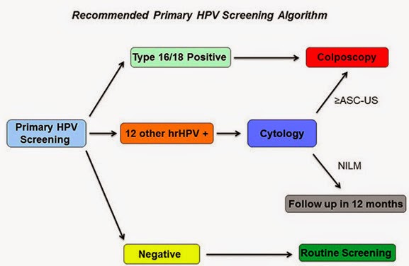
Under the new guidance if HPV test is positive for HPV types 16 or 18, colposcopy is the next step. If the HPV test is positive for one of 12 other HPV types, a Pap smear (cytology) is the next step. Credit: UAB
About 80 million U.S. women ages 25 to 65 should be screened periodically by their health care providers for cervical cancer. At present, the standard way to do that is a Pap smear alone, or co-testing using both a Pap smear and a human papillomavirus (HPV) test.

Today, the clinicians who care for those women are getting new interim guidance about the health advantages of instead using the HPV test alone as the primary screen to find cervical cancer or its precursors. Under the new guidance, the Pap smear, which dates back more than 80 years, would still be used for follow-up tests if an HPV test is positive. The Pap smear will still be used for primary screening of women under age 25.

The need for guidance about using the HPV test was triggered last April when the FDA approved one existing HPV test for use in primary cervical cancer screening. Today's guidance, written by a group of cervical cancer screening experts led by University of Alabama at Birmingham gynecologic oncologist Warner Huh, M.D., is being published simultaneously in the journals Gynecologic Oncology, Obstetrics & Gynecology, and the Journal of Lower Genital Tract Disease under the title "Use of Primary High Risk Human Papillomavirus Testing for Cervical Cancer Screening: Interim Clinical Guidance." Also published today in Gynecologic Oncology is the end-of-trial data of the Roche Diagnostics ATHENA HPV trial that enrolled more than 47,000 women in a longitudinal, three-year study of Roche's HPV test.

"Although FDA approval is critically important for introducing a new screening test or algorithm, providers ultimately rely on guidance or guidelines to help them make the best decisions for their patients and want to understand advantages, disadvantages and unknowns associated with a new screening approach," said Huh, who is a senior scientist for the UAB Comprehensive Cancer Center, Director of the UAB Division of Gynecologic Oncology, and is also a board member for both the American Society for Colposcopy and Cervical Pathology and the Society of Gynecologic Oncology.

Major conclusions The two major conclusions of the interim guidance panel are:

• "Because of equivalent or superior effectiveness," the paper says, "primary HPV screening can be considered as an alternative to current U.S. cytology-based (i.e., Pap smears) cervical cancer screening methods."

The authors note that the existing, previously published guidelines still recommend Pap smears alone, or co-testing with a Pap smear and an HPV test, for cervical cancer screening. However, those guidelines from 2011 predate more recent clinical studies of HPV testing that were analyzed in today's paper.

• Women who have a negative HPV test result from their primary screening have a greater reassurance of a very low risk for a future cervical cancer precursor lesion, as compared to women who have a negative Pap smear test in their primary screening. This lower rate of false negative results is a key benefit of the HPV screening. Overall, the panel said, "While there continue to be numerous practical and research questions, primary HPV testing has the potential to further reduce morbidity and mortality of cervical cancer in the U.S. 
However, what is most important is that women need to be screened with any strategy, as many women in the U.S. with cervical cancer are either unscreened or underscreened."

"The scientific evidence clearly demonstrates that primary HPV testing outperforms cytology or Pap as a screening test," said Huh. "This has been confirmed from numerous European and Canadian studies as well as the ATHENA trial. There are going to be fewer false negatives with HPV, and arguably, we have been using a less sensitive test for screening for a while now."

Huh added, "Pap smears miss a fair number of adenocarcinomas. "We don't want a test that will miss disease."

From the patient's point of view, the experience of getting an HPV test will be the same as getting a Pap smear. The difference is how the sample is then screened: Instead of a technician looking for abnormal cells (Pap), the HPV sample is put into an automated machine to detect HPV DNA.

Other questions The guidance also addresses four other questions that clinicians may have. 
1. How should one manage a positive HPV result? While data are still limited, the study group suggested a flowchart algorithm, as follows. If a woman is positive for HPV genotypes 16 or 18, which convey the greatest risk of developing cervical cancer precursor lesions in the next three years, she should be referred for a colposcopy (an illuminated, magnified examination of the cervix and other genital tissue for premalignant or malignant lesions). If a woman is positive for the 12 other lower-risk HPV genotypes, she should get a Pap smear; and if that Pap smear is also positive, she should then get a colposcopy. If her follow-up Pap smear is negative, she should be retested with another Pap smear in 12 months. This algorithm "achieves a reasonable balance of disease detection with the number of screening tests and colposcopies required to achieve that detection," the panel wrote.

2. What is the optimal interval for primary HPV screening?
Data are limited for determining the optimal screening interval, but the interval should be no sooner than every three years. There is no need to screen more frequently than every three years, since the cumulative occurrence of a cervical cancer precursor lesion called a CIN3+ during the three years after a negative HPV test was less than 1 percent.

3. At what age should one initiate primary HPV screening?
It should not begin before age 25. The study panel noted that about 30 percent of the CIN3+ cervical cancer precursor lesions in the ATHENA study occurred in women ages 25 to 29. A majority of women ages 25 to 29 who have CIN3+ have normal Pap smears. Another 37 percent of the CIN3+ lesions in the ATHENA study were found in women 30 to 39 years old. 
The panel did have some concerns that starting at age 25 -- even though it increases detection of disease -- would lead to too many colposcopies in women whose progression to cancer is uncommon.

4. How does primary HPV screening performance compare with co-testing?
The panel said that most of the reassurance of safety provided by a co-test (a Pap smear together with an HPV test) derives from the HPV test. Analysis of about 1 million women screened at Kaiser Permanente Northern California suggests that HPV screening with a three-year interval between negative tests is at least as effective as co-testing every five years. However, co-testing is still an appropriate and recommended screening strategy, Huh noted.

The future As the new advance of primary HPV screening enters into clinical practice, there will be a number of additional questions and concerns, the panel said. First, clinicians need to be aware that false negative tests will still occur -- that is, some women will still develop invasive cancer, even though their HPV tests were negative.

Second, at present there are four commercially available, FDA-approved HPV tests; but only one of them is FDA-approved for primary screening. While the panel hopes that there will be other tests that will be rigorously validated and approved for primary screening sometime in the near future, clinicians should not use a test that lacks a specific primary HPV screening indication.

Third, the panel noted a need for comparative effectiveness studies "that consider projected lifetime number of screening tests, colposcopies and follow-up visits," as well as direct cost comparisons between primary HPV testing vs. Pap smears and co-testing. Further information is also needed about the cancer risks if the interval between HPV tests is extended from three years to five years.

While today's guidance applies to women who receive regular screening for cervical cancer, 
the panel also noted the continuing need to identify women who are still unscreened or underscreened.

"One major aspect of cervical cancer prevention that needs to be discussed in light of screening is HPV vaccination," said Huh. "Particularly with the recent FDA approval of the new 9-valent HPV vaccine and evidence that the vaccine decreases HPV and disease prevalence, I have concerns that this will put an additional strain on the performance of cytology (i.e., Pap smear). We will need to look at other tests like HPV as a more appropriate screening test as disease rates decrease over time."

New approach aims to silence cancer 'survival genes'

Silencing the SIRT1 gene: Cancer cells before and after treatment in vitro. Non-cancerous cells (not shown) are unaffected. Credit: Image courtesy of University of York
Scientists at the University of York are working on a promising new approach for tackling colorectal cancer, the second most common cause of cancer-related death.

The new method works by silencing cancer 'survival genes' and could potentially combat cancer through the selective killing of colorectal cancer cells without adverse effects on normal, non-cancer cells.

Funded by York's Centre for Chronic Diseases and Disorders (C2D2), the project led by Professor Jo Milner from York's Department of Biology involved preliminary studies to establish the suitability of an ex vivo model for the future development of anti-cancer therapies for colorectal cancer using a technique called RNA interference.

The new approach builds on ground-breaking research by Professor Milner and her team at York more than a decade ago. This early work, funded by Yorkshire Cancer Research (YCR), used the newly-developed technique of RNA interference to successfully kill human cervical cancer cells grown in culture without causing damage to healthy cells.

Professor Milner explained: "When a mammalian cell elects to die it does so with great precision and without harming its neighbours. This process of 'programmed cell death' enables the continuous replacement of aging cells and also the sculpting of tissues and neuronal pathways.

"However, when this normal process of programmed cell death fails the continued abnormal growth of affected cells can lead to cancer. Some cancers develop following infection with a virus, such as human papilloma virus which causes human cervical cancer. Here the virus expresses specific viral genes that disrupt normal cellular control mechanisms resulting in abnormal cell proliferation and survival.

"Using RNA interference (RNAi) we first identified the viral gene responsible for the continued survival of cervical cancer cells. Then we established the feasibility of RNAi-based therapeutics for the selective killing of human cervical cancer cells growing in vitro."

Professor Milner and her team next studied cells from other cancer types, including colorectal cancer and breast cancer. Such cancers develop when the cell's internal control system fails due to damage to one or more of the regulatory genes.

Professor Milner said "We discovered that other genes, belonging to a group called stress-response genes, acquire a new pro-survival function during the process of cancerous transformation. Importantly, this acquired cancer-specific survival function operates under normal, physiological conditions. Silencing these cancer-specific survival genes by RNA interference causes the cancer cells to die while the survival of non-cancerous cells appears normal. This is in contrast to treating cancer by radiotherapy and/or genotoxic drugs -- these agents cause genotoxic stress and damage both cancer and normal cells and tissues in the body, resulting in unwanted adverse side effects for the patient."

For the work on colorectal cancer therapies to progress towards the clinic, the team has had to meet the challenge of modifying the agent siRNA. siRNA is the synthetic RNA molecule which is designed to silence a chosen gene by inducing RNA interference and selectively suppressing expression of that gene. However, siRNA is very unstable and is rapidly degraded when in contact with human tissues.

As reported in the journal Molecular Therapy, the team has now successfully met this challenge and converted the unstable siRNA molecule into a stable form without losing its ability and very high efficacy for targeted gene silencing. A novel siRNA/DNA has been shown to be resistant to degradation while retaining high efficacy and selectivity for target gene silencing when tested on human cancer cells grown in culture.

The next step will involve testing this novel therapeutic agent for cancer-specific cell killing using human tissue maintained ex vivo, using an experimental model which was validated in the course of the C2D2-funded research.

Professor Paul Kaye, Director of C2D2, said: "Professor Milner's team has now shown that ex vivo cultures of colorectal tumour material, derived from human patients, maintain cancer-related biochemistry over several days, and of sufficient time known to produce a killing effect with the novel siRNA/DNA in vitro. It is marvelous that C2D2 has been able to support this ground breaking research that has validated an ex vivo model that can be used to progress this novel therapeutic towards the clinic, and without the need for animal research."

Multiple allergic reactions traced to single protein

Written By Unknown on Friday, January 16, 2015 | 3:58 AM

This is a mast cell. Credit: Priyanka Pundir/University of Alberta
Johns Hopkins and University of Alberta researchers have identified a single protein as the root of painful and dangerous allergic reactions to a range of medications and other substances. If a new drug can be found that targets the problematic protein, they say, it could help smooth treatment for patients with conditions ranging from prostate cancer to diabetes to HIV. Their results appear in the journal Nature on Dec. 17.

Previous studies traced reactions such as pain, itching and rashes at the injection sites of many drugs to part of the immune system known as mast cells. When specialized receptors on the outside of mast cells detect warning signals known as antibodies, they spring into action, releasing histamine and other substances that spark inflammation and draw other immune cells into the area. Those antibodies are produced by other immune cells in response to bacteria, viruses or other perceived threats. However, "although many of these injection site reactions look like an allergic response, the strange thing about them is that no antibodies are produced," says Xinzhong Dong, Ph.D., an associate professor of neuroscience in the Institute for Basic Biomedical Sciences at the Johns Hopkins University School of Medicine.

To zero in on the cause of the reactions, Benjamin McNeil, Ph.D., a postdoctoral fellow in Dong's laboratory, first set out to find which mast cell receptor -- or receptors -- responded to the drugs in mice. Previous studies had identified a human receptor likely to be at fault in the allergic reactions; McNeil found a receptor in mice that, like the human receptor, is found only in mast cells. He then tested that receptor by putting it into lab-grown cells and found that they did react to medications that provoke mast cell response. He found similar results for the human receptor that previous studies had indicated was a likely culprit.

"It's fortunate that all of the drugs turn out to trigger a single receptor -- it makes that receptor an attractive drug target," McNeil says.

To find out whether eliminating the receptor really would eliminate the allergic reactions, the research team also disabled the gene for the suspect receptor in mice. These "knockout" mice did not have any of the drug allergy symptoms that their genetically normal counterparts displayed.

The researchers are now working to find compounds that could safely block the culprit receptor in humans, known as MRGPRX2. Such a drug would not prevent true allergic reactions, which produce antibodies, but only the pseudoallergic reactions triggered by MRGPRX2. Still, it could improve the lives of many patients, says McNeil, by lessening the drug side effects they currently endure. Medications that trigger MRGPRX2 include cancer drugs cetrorelix, leuprolide and octreotide; HIV drug sermorelin; fluoroquinolone antibiotics; and neuromuscular blocking drugs used to paralyze muscles during surgeries.

Dong's research group is also looking into the possibility that MRGPRX2 could be behind immune conditions such as rosacea and psoriasis that don't stem from medication use.

 
Support : Creating Website | Johny Template | Mas Template
Copyright © 2011. The planet wall - All Rights Reserved
Template Created by Easy Blogging Published by Mas Template
Proudly powered by Blogger