Latest Post
Showing posts with label BREAST CANCER. Show all posts

Cheap malaria drug could treat colorectal cancer effectively too, say experts

Written By Unknown on Sunday, January 18, 2015 | 4:34 PM

Artemisinin is isolated from the plant Artemisia annua also known as sweet wormwood. Credit: Image courtesy of University of St George's London
Medical experts say a common malaria drug could have a significant impact on colorectal cancer providing a cheap adjunct to current expensive chemotherapy.

A pilot study by researchers at St George's, University of London, has found the drug artesunate, which is a widely used anti-malaria medicine, had a promising effect on reducing the multiplication of tumour cells in colorectal cancer patients who were already going to have their cancer surgically removed.

Colorectal cancer (CRC) makes up about 10 percent of the annual 746,000 global cancer cases in men and 614,000 cases in women.

In the UK, 110 new cases are diagnosed daily, with older patients particularly at risk of death. Prognosis even with the best available treatments does not increase disease free or overall survival beyond 60 percent, five years after diagnosis.

Professor Sanjeev Krishna, an infectious disease expert at St George's who jointly-led the study, said: "There is therefore a continuing and urgent need to develop new, cheap, orally effective and safe colorectal cancer treatments.

"Our approach in this study was to take a close look at an existing drug that already had some anticancer properties in experimental settings, and to assess its safety and efficacy in patients.

"The results have been more than encouraging and can offer hopes of finding effective treatment options that are cheaper in the future."

"Larger clinical studies with artesunate that aim to provide well tolerated and convenient anticancer regimens should be implemented with urgency, and may provide an intervention where none is currently available, as well as synergistic benefits with current treatment regimens," added Professor Devinder Kumar, a leading expert in colorectal cancer at St George's and joint-lead of this study.

For most patients globally, access to advanced treatments is difficult as they are too expensive to be widely available, or associated with significant morbidity thereby further compromising their survival.

"In the St George's study, patients were examined and then were given either the anti-malaria drug artesunate or a placebo. After 42 months following surgery, there were six recurrences of cancer in the placebo group (of 12 patients) and one recurrence in an artesunate recipient (of 10 patients).The survival beyond two years in the artesunate group was estimated at 91% whilst surviving the first recurrence of cancer in the placebo group was only 57%.

This is the first randomized, double blind study to test the anti-CRC properties of oral artesunate. The anticancer properties of artemisinins have been seen in the laboratory previously but this is the first time their effect has been seen in patients in a rigorously designed study.

Researchers silence leading cancer-causing gene

Using bioluminescence, researchers showed that the novel molecule “KRAS silencing RNA” or "KRAS siRNA” (right) reduced the size of a tumor in mice. Researchers used a “non-KRAS silencing” molecule as the control (left) as a comparison. Credit: Image courtesy of University of North Carolina Health Care
Researchers from the UNC School of Medicine and colleagues at The University of Texas MD Anderson Cancer Center have developed a new approach to block the KRAS oncogene, one of the most frequently mutated genes in human cancer. The approach, led by Chad Pecot, MD, an assistant professor of medicine at UNC, offers another route to attack KRAS, which has proven to be an elusive and frustrating target for drug developers.

The new method relies upon a specifically sequenced type of small interfering RNA -- or siRNA. The findings, published in the journal Molecular Cancer Therapeutics, show that using a form of siRNA to halt KRAS not only dramatically stunted the growth of lung and colon cancers in cultured cells and mice but also stopped metastasis -- the main cause of cancer deaths.

"KRAS has been widely regarded as an undruggable protein, but we show that that's simply not the case," said Pecot, the study lead author and member of the UNC Lineberger Comprehensive Cancer Center.

KRAS is a signaling molecule -- a protein switch that triggers a cascade of molecular events that tell cells to grow and survive. Mutations in the KRAS gene create a switch that is perpetually "on," causing cells to divide uncontrollably. KRAS mutations are present in roughly 30 percent of human cancers, particularly lung, colon, pancreatic, and thyroid.

"It is the elephant in the room," Pecot said. "KRAS was one of the first cancer-causing genes ever discovered, and it was the obvious target to go after. People have been trying for decades to hit it, but they haven't had much luck."

Inhibiting KRAS signaling has been tricky because it lacks good pockets or crevices for small molecules and drugs to bind to. Some researchers have tried instead to target the proteins downstream in the KRAS signaling cascade, but those attempts have also had limited success.

Rather than try another conventional approach, Pecot decided to use a new genetic tool known as RNA interference -- or RNAi -- to destroy the KRAS protein before it fully forms. RNAi uses bits of synthetically engineered RNA -- the single-stranded molecule transcribed from DNA -- to silence specific genes. These bits of RNA bind to specific genetic messages called mRNA in the cell and direct enzymes to recognize the messages as enemies. In this context, the enzymes destroyed the genetic messages of KRAS mRNA so that KRAS can't be made. As a result, the cells don't grow, replicate, or move nearly as well.

RNAi has shown great promise in the treatment of liver diseases, viral infections, and cancers. To see if this approach could thwart the KRAS oncogene, Pecot and his colleagues first had to test different sequences of RNA to determine which one most effectively tagged KRAS for destruction. Of five RNA sequences, the researchers identified two candidates worthy to take into cancer models.

When they delivered these sequences into tissue culture cells, they found that the siRNAs destroyed more than 90 percent of the KRAS gene messages, significantly impairing the growth of cancer cell lines. The technique also led to marked reduction of two signaling molecules called pERK and pMEK, which lie downstream of KRAS and have been implicated in cancer cell proliferation and tumor growth.

Next, Pecot and his colleagues tested the siRNAs in mouse models of lung and colon cancer. They wrapped the sequences in protective lipid nanoparticles and delivered the siRNA solution into the mice. The researchers found that this treatment significantly slowed the growth of primary tumors. For example, tumors from colon cancer models that had been treated with the KRAS siRNAs were 69 percent smaller than tumors treated with control RNA sequences.

In addition, the researchers discovered that silencing KRAS stemmed the spread of cancer cells to other organs. The siRNA treatment reduced the number of these secondary malignant growths by about 80 percent in mice with lung cancer and to a similar degree in colon cancer models.

Pecot's findings come on the heels of two other papers using siRNAs to target KRAS, one from Frank McCormick's laboratory at the University of California at San Francisco and the other from Tyler Jacks' laboratory at the Massachusetts Institute of Technology. What sets the UNC study apart is that it demonstrates that this approach can be used to control the development of metastatic disease.

"Having all three papers come out at about the same time is encouraging because it means that KRAS is druggable if you use outside-the-box methods," Pecot said. "Now, we essentially have three platforms for targeting KRAS with siRNAs that may get to the clinic."

Pecot said the results, while promising, are just a first step in combating this cancer-causing gene. Ultimately, the siRNA sequences will have to be designed to specifically target the mutant form of KRAS without disrupting the normal form of the gene, which is necessary for maintaining normal growth in healthy cells.

Rewiring metabolism slows colorectal cancer growth

Many cancers have less MPC in them than normal adult tissues. Re-introducing MPC into cancer cells slows growth of tumors following injection into mice as compared to unmanipulated cells. Credit: Ralph DeBerardinis
Cancer is an unwanted experiment in progress. As the disease advances, tumor cells accumulate mutations, eventually arriving at ones that give them the insidious power to grow uncontrollably and spread. Distinguishing drivers of cancer from benign mutations open opportunities for developing targeted cancer therapies.

A University of Utah-led study reports that cancers select against a protein complex called the mitochondrial pyruvate carrier (MPC), and re-introduction of MPC in colon cancer cells impairs several properties of cancer, including growth. The research, which appears online on Oct. 30 in Molecular Cell, implicates changes in a key step in metabolism -- the way cellular fuel is utilized -- as an important driver of colon cancer that is also likely to be important in many other cancer settings.

Cancers appear to do whatever they can to get rid of MPC, a protein involved in carbohydrate metabolism, shows the study led by Jared Rutter, Ph.D., professor of biochemistry and Dee Glen and Ida W. Smith Endowed Chair for Cancer Research at the University of Utah. At least 18 types of cancers -- colon, brain, breast, and liver among them -- have significantly less MPC than normal adult cells. Some cancers simply delete a region of the genome that contains one of the MPC genes, others find different ways to dampen MPC expression. In fact, a survey of patient biopsies shows that the less MPC there is, the more aggressive the cancer becomes.

"Loss of MPC seems to be a biomarker for cancer aggressiveness and patient survival," said Rutter, also co-director of the Diabetes and Metabolism Center at the University of Utah, and co-leader of the Nuclear Control of Cell Growth and Differentiation Program at the Huntsman Cancer Institute. "That was our first clue that MPC might be important."

Even more striking, when Rutter's group reintroduced MPC into colon cancer cell lines, properties that define them as cancerous, reverted. The cells divide less frequently under certain conditions and decrease expression of stem cell markers, an early step frequently defining the potential to form tumors and spread. Further, the engineered cells are dramatically impaired in their ability to form tumors after injection into mice. Tumors containing cells with MPC were as small as one-fourth the size of tumors made from cells without the protein complex.

"We think these results show that elimination of MPC is an early and important step in development of cancer," said John Schell, who is co-first author with Kristofor Olson, both M.D.-Ph.D. students at the University of Utah. "Finding the stem cell connection was probably the most exciting part for us, and is something we'll pursue further to understand how loss of MPC changes cell behavior."

The role of MPC in the normal cell, and what loss of MPC does to a cancer cell, addresses an observation first made nearly one century ago. Nobel Prize-wining biochemist Otto Warburg noted that cancer cells change their metabolism to support uncontrolled growth and proliferation. Scientists later found the way in which the metabolite pyruvate is processed is key to these metabolic changes. In normal adult cells, pyruvate enters the mitochondria, the cell's powerhouse, and fuels energy production. In cancer, pyruvate is diverted from the mitochondria to an alternative metabolic pathway that makes cell-building material.

Scientists had long suspected the so-called Warburg effect seen in cancer was contingent upon controlling entry of pyruvate into the mitochondria. But there was no way to directly test the idea until two years ago, when Rutter's group and others identified MPC as pyruvate's doorway to the mitochondria. The current report in Molecular Cell shows that cancer cells shut that door by repressing MPC, and that experimentally re-opening the door by re-introducing MPC not only inhibits cancer growth, but also redirects pyruvate to the metabolic pathway used in normal cells. In other words, MPC counteracts the Warburg effect.

"This makes sense because MPC is a pinch point in metabolism," said Rutter. "Our work, taken together with that from many other laboratories, shows that most cancer cells are completely reliant on this unusual metabolism known as the Warburg effect."

Understanding the Warburg effect has been an area of intense interest in recent years because of the potential to translate those discoveries into new cancer therapeutics. "We think this information can be used to design therapies that are specifically toxic to cancer cells," said Rutter.

What makes pancreatic cancer so aggressive? New study sheds light

“We know that patients with the earliest stage of pancreatic cancer have a survival rate of only 30 percent. This suggests that even in that very early stage of invasive cancer there are already cells that have spread to distant parts of the body,” says study author Diane M. Simeone, M.D. Credit: Image courtesy of University of Michigan Health System
New research from the University of Michigan Comprehensive Cancer Center helps explain why pancreatic cancer is so lethal, with fewer than a third of patients surviving even early stage disease.

The researchers found a gene known to be involved in nearly 90 percent of pancreatic cancers promotes cancer growth and spread. The gene, ATDC, plays a key role in how a tumor progresses from a preinvasive state to an invasive cancer to metastatic cancer.

"We know that patients with the earliest stage of pancreatic cancer have a survival rate of only 30 percent. This suggests that even in that very early stage of invasive cancer there are already cells that have spread to distant parts of the body," says study author Diane M. Simeone, M.D., director of the Pancreatic Cancer Center at the University of Michigan Comprehensive Cancer Center.

"This study sheds important light on what it is about pancreatic cancer that makes it so aggressive early in the game," she adds. The study appears Jan. 15 in Genes and Development.

Researchers used a mouse model to replicate pancreatic cancer as it appears in humans. They also studied pancreatic cancer tissue samples and samples of pre-invasive pancreatic lesions. They found ATDC was expressed in a subset of the pre-invasive cells and played a role in the development of pancreatic cancer stem cells, the small number of cells in a tumor that fuel its growth and spread. This suggests that ATDC promotes a tumor's invasiveness and spread early in the course of disease.

The researchers suspect that ATDC may be a potent drug target. No drugs currently exist to target this pathway in part because researchers do not understand the crystal structure of the protein. Simeone's team, working with the University of Michigan Center for Structural Biology has made crystals of the protein and begun to create a three-dimensional structure that they can use as a model for drug development.

Preliminary data suggests that ATDC may also play a role in other cancer types, including bladder, ovarian, colorectal and lung cancers and multiple myeloma. But, Simeone notes, it's particularly critical to find new treatment options for pancreatic cancer. About 46,400 Americans will be diagnosed with pancreatic cancer this year, and more than 39,000 will die of the disease. Pancreatic cancer is expected to become the second-leading cause of cancer death in the United States by 2030.

3-D culture system for pancreatic cancer has potential to change therapeutic approaches

A team of researchers has developed a method to grow pancreatic tissue in a three-dimensional culture system, called organoids. The scientists are able to use tissue not only from laboratory mouse models, but also from human patients. The technology promises to change the way pancreatic cancer research is done, offering a path to personalized treatment approaches in the future. Credit: D. Tuveson/ Cold Spring Harbor Laboratory
Cold Spring Harbor and Bethpage, N.Y. -- Pancreatic cancer is one of the most deadly forms of cancer, with only 6 percent of patients surviving five years after diagnosis. Today, Cold Spring Harbor Laboratory (CSHL) and The Lustgarten Foundation jointly announce the development of a new model system to grow both normal and cancerous pancreatic cells in the laboratory. Their work offers the potential to change the way pancreatic cancer research is done, allowing scientists to interrogate the pathways driving this devastating disease while searching for new drug targets.

In work published in Cell, the research team describes a three-dimensional "organoid" culture system for pancreatic cancer. Co-led by David Tuveson, CSHL Professor and Director of Research for The Lustgarten Foundation, and Hans Clevers, Professor and Director of the Hubrecht Institute and President of the Royal Netherlands Academy of Arts and Sciences, the team developed a method to grow pancreatic tissue not only from laboratory mouse models, but also from human patient tissue, offering a path to personalized treatment approaches in the future.

All cancer research relies on a steady supply of cells -- both normal and cancerous -- that can be grown in the laboratory. By comparing normal cells to cancer cells, scientists can then identify the changes that lead to disease. However, both types of pancreatic cells have been extremely difficult to culture in the laboratory.

Furthermore, the normal ductal cells that are able to develop into pancreatic cancer represent about 10 percent of the cells in the pancreas, complicating efforts to pinpoint the changes that occur as the tumor develops. Until now, scientists have been entirely unable to culture human normal ductal pancreatic cells under standard laboratory conditions. 

Because of these limitations, most pancreatic cancer research relies on genetically engineered mouse models of the disease, which can take up to one year to generate. "With this development, we are now able to culture both mouse and human organoids, providing a very powerful tool in our fight against pancreatic cancer," explains Tuveson.

The organoids are entirely made up of ductal cells, eliminating the surrounding cell types that often contaminate samples from the pancreas. They grow as hollow spheres within a complex gel-like substance filled with growth-inducing factors and connecting fibers. Once they have grown to a sufficient size, the organoids can be transplanted back into mice, where they fully recapitulate pancreatic cancer. "We now have a model for each stage in the progression of the disease," says Chang-Il Hwang, Ph.D., one of the lead authors working in The Lustgarten Foundation's Pancreatic Cancer Research Lab at CSHL directed by Dr. Tuveson.

Traditionally, cancer cells are isolated during surgery or autopsies. Unfortunately, approximately 85 percent of cancer patients are ineligible for surgery at the time of diagnosis, either because the tumor is entwined in critical vasculature or the disease has progressed too far. Researchers therefore have had limited access to patient samples. The new research provides a way for scientists to grow organoids from biopsy material, which is comparatively easy to obtain. "Biopsies are the standard for diagnosis," says Dannielle Engle, Ph.D., also a lead author on the paper. "We can now rapidly generate organoids from any patient, which offers us the potential to study the disease in a much wider population."

The team is now working to create a repository of pancreatic tumor samples, coordinating with the National Cancer Institute. "We hope to make this available to the entire pancreatic cancer research community," says Tuveson. Additionally, Lindsey Baker, Ph.D., another lead author of the paper, has started holding an "organoid school" for other researchers, and has already taught six laboratories from around the world this technique.

'Sugar-coated' microcapsule eliminates toxic punch of experimental anti-cancer drug

3BrPA (red) is illustrated encased in a sugar-based microshell. Credit: Jean-Francois Geschwind, Johns Hopkins
Johns Hopkins researchers have developed a sugar-based molecular microcapsule that eliminates the toxicity of an anticancer agent developed a decade ago at Johns Hopkins, called 3-bromopyruvate, or 3BrPA, in studies of mice with implants of human pancreatic cancer tissue. The encapsulated drug packed a potent anticancer punch, stopping the progression of tumors in the mice, but without the usual toxic effects.

"We developed 3BrPA to target a hallmark of cancer cells, namely their increased dependency on glucose compared with normal cells. But the nonencapsulated drug is toxic to healthy tissues and inactivated as it navigates through the blood, so finding a way to encapsulate the drug and protect normal tissues extends its promise in many cancers as it homes in on tumor cells," says Jean-Francois Geschwind, M.D., chief of the Division of Interventional Radiology at Johns Hopkins Medicine.

The Johns Hopkins team used a microshell made of a sugar-based polymer called cyclodextrin to protect the 3BrPA drug molecules from disintegrating early and to guard healthy tissue from the drug's toxic effects, such as weight loss, hypothermia and lethal hypoglycemic shock.

Geschwind, a professor in the Russell H. Morgan Department of Radiology and Radiological Science at the Johns Hopkins University School of Medicine and its Kimmel Cancer Center, and others at Johns Hopkins have been studying the experimental drug as a cancer treatment for over a decade because of its ability to block a key metabolic pathway of cancer cells.

Most cancer cells, he explains, rely on the use of glucose to thrive, a process known as the Warburg effect, for Otto Heinrich Warburg, who was awarded the Nobel Prize in Physiology for the discovery in 1931. By using the same cellular channels that funnel glucose into a cancer cell, 3BrPA can travel inside the cancer cell and block its glucose metabolic pathway, Geschwind says.

However, animal studies have shown that in its free, nonencapsulated state, the drug is very toxic, says Geschwind.

The toxicity associated with the free-form version of the drug, he says, has prevented physicians from using the drug as a systemic treatment in people, one that can travel throughout the whole body.

In a report about their study published online Oct. 17 in Clinical Cancer Research, the researchers described minimal or zero tumor progression in mice treated with the microencapsulated 3BrPA. By contrast, a signal of tumor activity increased sixty-fold in mice treated with the widely used chemotherapy drug gemcitabine. Activity increased 140-fold in mice who received the drug without encapsulation.

Specifically, daily injections of nonencapsulated 3BrPA were highly toxic to the animals, as only 28 percent of the animals survived the 28-day treatment. All of the mice who received the encapsulated drug survived to the end of the study.

Geschwind says the "extremely promising results" of the study make the encapsulated drug a good candidate for clinical trials, particularly for patients with pancreatic ductal adenocarcinoma. These cancers rank as the fourth most common cause of cancer-related deaths in the world, with a five-year survival rate of less than 5 percent. In the mouse studies, the encapsulated medication also reduced the metastatic spread of pancreatic cancer cells.

New combination therapy developed for multiple myeloma

This is Steven Grant, M.D., Shirley Carter Olsson and Sture Gordon Olsson Chair in Cancer Research, associate director for translational research and program co-leader of Developmental Therapeutics at VCU Massey Cancer Center. Credit: VCU Massey Cancer Center
Each year, more than 25,000 Americans are diagnosed with multiple myeloma, a form of blood cancer that often develops resistance to therapies. However, researchers at Virginia Commonwealth University Massey Cancer Center are reporting promising results from laboratory experiments testing a new combination therapy that could potentially overcome the resistance hurdle.

While several drugs are effective against multiple myeloma, including the proteasome inhibitor bortezomib, multiple myeloma cells are often able to survive by increasing the production of a protein known as Mcl-1. Mcl-1 regulates a number of processes that promote cell survival and has been implicated in resistance to anti-myeloma drugs that were initially effective. However, a team of researchers led by Xin-Yan Pei, M.D., Ph.D., and Steven Grant, M.D., recently published the findings of a study in the journal PLoS ONE demonstrating that a novel drug combination both reduces Mcl-1 expression and disrupts its interactions with other proteins to effectively kill multiple myeloma cells. The therapy combines a type of drug known as a Chk1 inhibitor with another called a MEK inhibitor. Chk1 inhibitors prevent cells from arresting in stages of the cell cycle that facilitate the repair of DNA damage, while MEK inhibitors prevent cells from activating a variety of proteins that regulate DNA repair processes while promoting the accumulation of pro-death proteins.

"This research builds on our previous studies that showed exposing multiple myeloma and leukemia cells to Chk1 inhibitors activated a protective response through the Ras/MEK/ERK signaling pathway," says Pei, instructor in the Department of Internal Medicine at the VCU School of Medicine. "By combining a Chk1 inhibitor with a MEK inhibitor, we have developed one of only a limited number of strategies shown to circumvent therapeutic resistance caused by high expressions of Mcl-1."

In laboratory experiments, the scientists enforced overexpression of Mcl-1 in human multiple myeloma cells. They found that this caused the cells to become highly resistant to bortezomib, but it failed to protect them from the Chk1/MEK inhibitor regimen. 
Additionally, the combination therapy was able to completely overcome resistance due to microenvironmental factors associated with increased expression of Mcl-1. A cell's microenvironment consists of surrounding cells and the fluids in which they reside, and the communication between cancer cells and their surrounding cells can significantly impact resistance. Mcl-1 plays a key role in this communication by facilitating events that promote cancer cell survival.

"Not only was the combination therapy effective against multiple myeloma cells, it notably did not harm normal bone marrow cells, raising the possibility of therapeutic selectivity," says Grant, the study's lead investigator and Shirley Carter Olsson and Sture Gordon Olsson Chair in Cancer Research, associate director for translational research and program co-leader of Developmental Therapeutics at VCU Massey Cancer Center. "We are hopeful that this research will lead to better therapies for multiple myeloma, and help make current therapies more effective by overcoming resistance caused by Mcl-1."

The researchers have started initial discussions with clinical investigators and drug manufacturers with hopes of developing a clinical trial testing a combination of Chk1 and MEK inhibitors in patients with refractory multiple myeloma. It is too early to estimate when the trial will open.

Powerful new system for classifying tumors revealed

This diagram illustrates how tumors with different tissues of origin were reclassified on the basis of molecular analyses. Credit: Zhong Chen, NIH/NIDCD
Cancers are classified primarily on the basis of where in the body the disease originates, as in lung cancer or breast cancer. According to a new study, however, one in ten cancer patients would be classified differently using a new classification system based on molecular subtypes instead of the current tissue-of-origin system. This reclassification could lead to different therapeutic options for those patients, scientists reported in a paper published August 7 in Cell.

"It's only ten percent that were classified differently, but it matters a lot if you're one of those patients," said senior author Josh Stuart, a professor of biomolecular engineering at UC Santa Cruz.

Stuart helped organize the study as part of the Pan-Cancer Initiative of the Cancer Genome Atlas (TCGA) project. A large team of researchers from multiple institutions performed a comprehensive analysis of molecular data from thousands of patients representing 12 different types of cancer. This was the most comprehensive and diverse collection of tumors ever analyzed by systematic genomic methods. Each tumor type was characterized using six different "platforms" or methods of molecular analysis--mostly genomic platforms such as DNA and RNA sequencing, plus a protein expression analysis.

The research team used statistical analyses of the molecular data to divide the tumors into groups or "clusters," first analyzing the data from each platform separately and then combining them in an integrated cross-platform analysis developed by co-first author Katherine Hoadley of the University of North Carolina. All six platforms as well as the integrated analysis converged on the same divisions of the cancers into 11 major subtypes. 
Five of those subtypes were nearly identical to their tissue-of-origin counterparts. But some tissue-of-origin categories split into several different molecular subtypes, and some subtypes encompass tumors with several different tissues of origin.

Bladder cancer was a particularly interesting group, because it split into seven different clusters, with most samples falling into one of three subtypes. One subtype was bladder cancer only, but some bladder cancers clustered with lung adenocarcinomas, and others with a subtype called 'squamous-like' that includes some lung cancers, some head-and-neck cancers, and some bladder cancers.

"If you look at survival rates, the bladder cancers that clustered with other tumor types had a worse prognosis. So this is not just an academic exercise," Stuart said.

Other findings from the study reconfirmed cancer subtypes that were already recognized, such as the different subtypes of breast cancer based on well-characterized biomarkers. The findings provide a more refined, quantitative picture of the differences between breast cancer subtypes, Stuart said. For example, the results reinforce the idea that 'basal-like' breast cancers are a unique tumor type. "Basal-like breast cancers are as different from luminal breast cancers as they are from lung cancers," he said.

The fact that all six platforms for molecular analysis identified the same set of subtypes, both individually and in multi-platform analyses, is an important result, Stuart noted. Not only does it give the researchers confidence in the subtypes they identified, it also means that different kinds of data can be used to classify a tumor.

"We can now say what the telltale signatures of the subtypes are, so you can classify a patient's tumor just based on the gene expression data, or just based on mutation data, if that's what you have," Stuart said. "Having a molecular map like this could help get a patient into the right clinical trial."

Although follow-up studies are needed to validate the findings, this new analysis lays the groundwork for classifying tumors into molecularly defined subtypes. The new classification scheme could be used to enroll patients in clinical trials and could lead to different treatment options based on molecular subtypes.

According to Stuart, the percentage of tumors that are reclassified based on molecular signatures is likely to grow as more samples and tumor types are included in the analysis (the next major Pan-Cancer analysis will include 21 tumor types). Coauthor Christopher Benz, an oncologist at the Buck Institute for Research on Aging and UC San Francisco, noted that the 10 percent reclassification rate in the current study is likely an underestimate due to the unequal representation of different tumors. "If our study had included as many bladder cancers as breast cancers, for example, we would have reclassified 30 percent," Benz said.

The researchers reported that each molecular subtype may reflect tumors arising from distinct cell types. For example, the data showed a marked difference between cancers of epithelial and non-epithelial origins. "We think the subtypes reflect primarily the cell of origin. Another factor is the nature of the genomic lesion, and third is the microenvironment of the cell and how surrounding cells influence it," Stuart said. "We are disentangling the signals from these different factors so we can gauge each one for its prognostic power."

The study involved an enormous amount of molecular and clinical data, which was managed by data coordinator Kyle Ellrott, a software developer in Stuart's lab at UC Santa Cruz. The data sets and results have been made available to other researchers through the Synapse web site (http://www.synapse.org). Stuart worked with the bioinformatics company Sage Bionetworks to create Synapse as a data repository for the Pan-Cancer Initiative.

"It's a huge amount of information, and all the data is available as programmable data sets that other researchers can use to do further analysis," Stuart said. "The scale of this project is hard to imagine. All of the data that the TCGA project has been churning out got funneled into this paper, and it's giving us an unbiased look at what the data have to tell us about cancer."

New recommendation for cervical cancer screening, using HPV test alone

Under the new guidance if HPV test is positive for HPV types 16 or 18, colposcopy is the next step. If the HPV test is positive for one of 12 other HPV types, a Pap smear (cytology) is the next step. Credit: UAB
About 80 million U.S. women ages 25 to 65 should be screened periodically by their health care providers for cervical cancer. At present, the standard way to do that is a Pap smear alone, or co-testing using both a Pap smear and a human papillomavirus (HPV) test.

Today, the clinicians who care for those women are getting new interim guidance about the health advantages of instead using the HPV test alone as the primary screen to find cervical cancer or its precursors. Under the new guidance, the Pap smear, which dates back more than 80 years, would still be used for follow-up tests if an HPV test is positive. The Pap smear will still be used for primary screening of women under age 25.

The need for guidance about using the HPV test was triggered last April when the FDA approved one existing HPV test for use in primary cervical cancer screening. Today's guidance, written by a group of cervical cancer screening experts led by University of Alabama at Birmingham gynecologic oncologist Warner Huh, M.D., is being published simultaneously in the journals Gynecologic Oncology, Obstetrics & Gynecology, and the Journal of Lower Genital Tract Disease under the title "Use of Primary High Risk Human Papillomavirus Testing for Cervical Cancer Screening: Interim Clinical Guidance." Also published today in Gynecologic Oncology is the end-of-trial data of the Roche Diagnostics ATHENA HPV trial that enrolled more than 47,000 women in a longitudinal, three-year study of Roche's HPV test.

"Although FDA approval is critically important for introducing a new screening test or algorithm, providers ultimately rely on guidance or guidelines to help them make the best decisions for their patients and want to understand advantages, disadvantages and unknowns associated with a new screening approach," said Huh, who is a senior scientist for the UAB Comprehensive Cancer Center, Director of the UAB Division of Gynecologic Oncology, and is also a board member for both the American Society for Colposcopy and Cervical Pathology and the Society of Gynecologic Oncology.

Major conclusions The two major conclusions of the interim guidance panel are:

• "Because of equivalent or superior effectiveness," the paper says, "primary HPV screening can be considered as an alternative to current U.S. cytology-based (i.e., Pap smears) cervical cancer screening methods."

The authors note that the existing, previously published guidelines still recommend Pap smears alone, or co-testing with a Pap smear and an HPV test, for cervical cancer screening. However, those guidelines from 2011 predate more recent clinical studies of HPV testing that were analyzed in today's paper.

• Women who have a negative HPV test result from their primary screening have a greater reassurance of a very low risk for a future cervical cancer precursor lesion, as compared to women who have a negative Pap smear test in their primary screening. This lower rate of false negative results is a key benefit of the HPV screening. Overall, the panel said, "While there continue to be numerous practical and research questions, primary HPV testing has the potential to further reduce morbidity and mortality of cervical cancer in the U.S. 
However, what is most important is that women need to be screened with any strategy, as many women in the U.S. with cervical cancer are either unscreened or underscreened."

"The scientific evidence clearly demonstrates that primary HPV testing outperforms cytology or Pap as a screening test," said Huh. "This has been confirmed from numerous European and Canadian studies as well as the ATHENA trial. There are going to be fewer false negatives with HPV, and arguably, we have been using a less sensitive test for screening for a while now."

Huh added, "Pap smears miss a fair number of adenocarcinomas. "We don't want a test that will miss disease."

From the patient's point of view, the experience of getting an HPV test will be the same as getting a Pap smear. The difference is how the sample is then screened: Instead of a technician looking for abnormal cells (Pap), the HPV sample is put into an automated machine to detect HPV DNA.

Other questions The guidance also addresses four other questions that clinicians may have. 
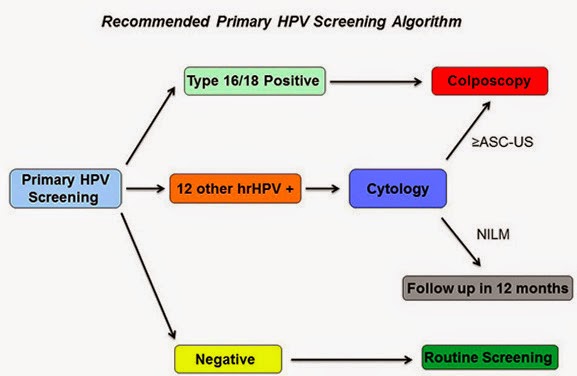
1. How should one manage a positive HPV result? While data are still limited, the study group suggested a flowchart algorithm, as follows. If a woman is positive for HPV genotypes 16 or 18, which convey the greatest risk of developing cervical cancer precursor lesions in the next three years, she should be referred for a colposcopy (an illuminated, magnified examination of the cervix and other genital tissue for premalignant or malignant lesions). If a woman is positive for the 12 other lower-risk HPV genotypes, she should get a Pap smear; and if that Pap smear is also positive, she should then get a colposcopy. If her follow-up Pap smear is negative, she should be retested with another Pap smear in 12 months. This algorithm "achieves a reasonable balance of disease detection with the number of screening tests and colposcopies required to achieve that detection," the panel wrote.

2. What is the optimal interval for primary HPV screening?
Data are limited for determining the optimal screening interval, but the interval should be no sooner than every three years. There is no need to screen more frequently than every three years, since the cumulative occurrence of a cervical cancer precursor lesion called a CIN3+ during the three years after a negative HPV test was less than 1 percent.

3. At what age should one initiate primary HPV screening?
It should not begin before age 25. The study panel noted that about 30 percent of the CIN3+ cervical cancer precursor lesions in the ATHENA study occurred in women ages 25 to 29. A majority of women ages 25 to 29 who have CIN3+ have normal Pap smears. Another 37 percent of the CIN3+ lesions in the ATHENA study were found in women 30 to 39 years old. 
The panel did have some concerns that starting at age 25 -- even though it increases detection of disease -- would lead to too many colposcopies in women whose progression to cancer is uncommon.

4. How does primary HPV screening performance compare with co-testing?
The panel said that most of the reassurance of safety provided by a co-test (a Pap smear together with an HPV test) derives from the HPV test. Analysis of about 1 million women screened at Kaiser Permanente Northern California suggests that HPV screening with a three-year interval between negative tests is at least as effective as co-testing every five years. However, co-testing is still an appropriate and recommended screening strategy, Huh noted.

The future As the new advance of primary HPV screening enters into clinical practice, there will be a number of additional questions and concerns, the panel said. First, clinicians need to be aware that false negative tests will still occur -- that is, some women will still develop invasive cancer, even though their HPV tests were negative.

Second, at present there are four commercially available, FDA-approved HPV tests; but only one of them is FDA-approved for primary screening. While the panel hopes that there will be other tests that will be rigorously validated and approved for primary screening sometime in the near future, clinicians should not use a test that lacks a specific primary HPV screening indication.

Third, the panel noted a need for comparative effectiveness studies "that consider projected lifetime number of screening tests, colposcopies and follow-up visits," as well as direct cost comparisons between primary HPV testing vs. Pap smears and co-testing. Further information is also needed about the cancer risks if the interval between HPV tests is extended from three years to five years.

While today's guidance applies to women who receive regular screening for cervical cancer, 
the panel also noted the continuing need to identify women who are still unscreened or underscreened.

"One major aspect of cervical cancer prevention that needs to be discussed in light of screening is HPV vaccination," said Huh. "Particularly with the recent FDA approval of the new 9-valent HPV vaccine and evidence that the vaccine decreases HPV and disease prevalence, I have concerns that this will put an additional strain on the performance of cytology (i.e., Pap smear). We will need to look at other tests like HPV as a more appropriate screening test as disease rates decrease over time."

Mushroom extract, AHCC, helpful in treating HPV

Judith A. Smith, Pharm.D. Credit: Image courtesy of University of Texas Health Science Center at Houston
A Japanese mushroom extract appears to be effective for the eradication of human papillomavirus (HPV), according to a pilot clinical trial at The University of Texas Health Science Center at Houston (UTHealth) Medical School.

The results were presented at the 11th International Conference of the Society for Integrative Oncology in Houston today by principal investigator Judith A. Smith, Pharm.D., associate professor in the Department of Obstetrics, Gynecology and Reproductive Sciences at the UTHealth Medical School.

Ten HPV-positive women were treated orally with the extract, AHCC (active hexose correlated compound) once daily for up to six months. Five achieved a negative HPV test result -- three with confirmed eradication after stopping AHCC -- with the remaining two responders continuing on the study.

Currently, there is no effective medicine or supplement to treat HPV, which is associated with more than 99 percent of cervical cancer cases. According to the Centers for Disease Control and Prevention, several other cancers are related to HPV, including 95 percent of anal cancer, 60 percent of oropharyngeal, 65 percent of vaginal cancer, 50 percent of vulvar cancer and 35 percent of penile cancer.

AHCC is a readily available nutritional supplement that works to improve the innate immune system. Human and preclinical studies have shown that AHCC increases the number and/or activity of Natural Killer (NK) cells, dendritic cells and cytokines, which help the body fight off infections and block tumor growth.

"The results are very encouraging," Smith said. "We were able to determine that at least three months of treatment is necessary but some need to extend that to six months. Since AHCC is a nutritional supplement with no side effects and other immune modulating benefits, we will be planning on using six months of treatment in our phase II clinical study to have consistent study treatment plan. This confirms our earlier preclinical research."

Smith is director of UTHealth's Women's Health Integrative Medicine Research Team, which focuses on the safe and effective use of nutritional and herbal supplements with pharmacologic modalities as it relates to women's health and cancer.

This research is proceeding to a randomized, double-blind, placebo-controlled Phase II clinical trial which has just begun at UTHealth, Smith said. For more information on enrolling in the trial, go to http://go.uth.edu/judithresearch.

New approach aims to silence cancer 'survival genes'

Silencing the SIRT1 gene: Cancer cells before and after treatment in vitro. Non-cancerous cells (not shown) are unaffected. Credit: Image courtesy of University of York
Scientists at the University of York are working on a promising new approach for tackling colorectal cancer, the second most common cause of cancer-related death.

The new method works by silencing cancer 'survival genes' and could potentially combat cancer through the selective killing of colorectal cancer cells without adverse effects on normal, non-cancer cells.

Funded by York's Centre for Chronic Diseases and Disorders (C2D2), the project led by Professor Jo Milner from York's Department of Biology involved preliminary studies to establish the suitability of an ex vivo model for the future development of anti-cancer therapies for colorectal cancer using a technique called RNA interference.

The new approach builds on ground-breaking research by Professor Milner and her team at York more than a decade ago. This early work, funded by Yorkshire Cancer Research (YCR), used the newly-developed technique of RNA interference to successfully kill human cervical cancer cells grown in culture without causing damage to healthy cells.

Professor Milner explained: "When a mammalian cell elects to die it does so with great precision and without harming its neighbours. This process of 'programmed cell death' enables the continuous replacement of aging cells and also the sculpting of tissues and neuronal pathways.

"However, when this normal process of programmed cell death fails the continued abnormal growth of affected cells can lead to cancer. Some cancers develop following infection with a virus, such as human papilloma virus which causes human cervical cancer. Here the virus expresses specific viral genes that disrupt normal cellular control mechanisms resulting in abnormal cell proliferation and survival.

"Using RNA interference (RNAi) we first identified the viral gene responsible for the continued survival of cervical cancer cells. Then we established the feasibility of RNAi-based therapeutics for the selective killing of human cervical cancer cells growing in vitro."

Professor Milner and her team next studied cells from other cancer types, including colorectal cancer and breast cancer. Such cancers develop when the cell's internal control system fails due to damage to one or more of the regulatory genes.

Professor Milner said "We discovered that other genes, belonging to a group called stress-response genes, acquire a new pro-survival function during the process of cancerous transformation. Importantly, this acquired cancer-specific survival function operates under normal, physiological conditions. Silencing these cancer-specific survival genes by RNA interference causes the cancer cells to die while the survival of non-cancerous cells appears normal. This is in contrast to treating cancer by radiotherapy and/or genotoxic drugs -- these agents cause genotoxic stress and damage both cancer and normal cells and tissues in the body, resulting in unwanted adverse side effects for the patient."

For the work on colorectal cancer therapies to progress towards the clinic, the team has had to meet the challenge of modifying the agent siRNA. siRNA is the synthetic RNA molecule which is designed to silence a chosen gene by inducing RNA interference and selectively suppressing expression of that gene. However, siRNA is very unstable and is rapidly degraded when in contact with human tissues.

As reported in the journal Molecular Therapy, the team has now successfully met this challenge and converted the unstable siRNA molecule into a stable form without losing its ability and very high efficacy for targeted gene silencing. A novel siRNA/DNA has been shown to be resistant to degradation while retaining high efficacy and selectivity for target gene silencing when tested on human cancer cells grown in culture.

The next step will involve testing this novel therapeutic agent for cancer-specific cell killing using human tissue maintained ex vivo, using an experimental model which was validated in the course of the C2D2-funded research.

Professor Paul Kaye, Director of C2D2, said: "Professor Milner's team has now shown that ex vivo cultures of colorectal tumour material, derived from human patients, maintain cancer-related biochemistry over several days, and of sufficient time known to produce a killing effect with the novel siRNA/DNA in vitro. It is marvelous that C2D2 has been able to support this ground breaking research that has validated an ex vivo model that can be used to progress this novel therapeutic towards the clinic, and without the need for animal research."

Artificial light, biological clock disruptions, increase breast cancer risk, study finds

Written By Unknown on Friday, January 16, 2015 | 3:44 AM

Employment as a flight attendant has been found to be related to an increased risk of breast cancer. Credit: © dmitrimaruta / Fotolia
The disruption of a person's circadian rhythm -- their 24-hour biological clock -- has been linked to an increased risk of breast cancer, according to new University of Georgia research. The culprit, in this study in particular, is artificial light.

"Exposure to artificial light leads to a significantly higher risk for developing breast cancer," said Chunla He, a biostatistics graduate student in the UGA College of Public Health. "To decrease the use of artificial light, people should avoid working at night and implement earlier bed times."

Her research, published in the International Archives of Occupational and Environmental Health, examined key studies that included risk factors for developing breast cancer.

"A large body of related research about circadian rhythms and breast cancer exists," He said. 
"However, these studies are inconsistent and have a variety of limitations."

Under the mentorship of Sara Wagner Robb, assistant professor of epidemiology in the College of Public Health, He turned to previous studies to see what the research revealed.

In addition to other relevant exposures, He examined studies on breast cancer and flight attendants, who typically work both day and night shifts. The flight attendants represented a group of workers particularly susceptible to disrupted circadian rhythms, which are heavily influenced by light.

In her analysis, He found that employment as a flight attendant was related to an increased risk of breast cancer.

"People naturally secrete the hormone melatonin, which helps to regulate the circadian rhythm," He said. "When the sleep-wake cycle is disrupted by artificial light, melatonin secretion is adversely affected."

Robb recommends spreading this information to shift workers so they understand the harms in disrupting their circadian rhythms.

"Individuals engaging in this type of work should be aware of these risks and may make efforts to adjust their circadian rhythms," she said. "Although additional studies are certainly needed, scientists are becoming increasingly aware of the health risks associated with night workers and others exposed to circadian-disrupting behaviors."

Robb and He also advise that future research needs to examine social constraints -- which may foster disruption of circadian rhythms -- on shift workers. Additionally, shift workers should contact their primary care physicians for personalized treatment and options.

"This information tells us the harm in disrupting our natural cycle," He said. "With this new analysis, we must be cautious in our exposure to artificial light."

The article was co-authored by Sonia Taj Anand, a former graduate student in the College of Public Health; Mark H. Ebell, professor of epidemiology; and John E. Vena, Medical University of South Carolina.

Looks really can kill you: Protect yourself against skin cancer

Written By Unknown on Thursday, January 15, 2015 | 7:09 PM

Credit: Image courtesy of Seattle Cancer Care Alliance
It only takes a few bad sunburns or trips to the tanning bed to put someone at risk for melanoma. Skin cancer is the most common type of cancer in the United States and when left untreated, melanoma is the most dangerous and aggressive form. It accounts for more than 9,000 of the 12,000-plus skin cancer deaths each year. In observance of May's Melanoma and Skin Cancer Awareness Month Seattle Cancer Care Alliance (SCCA) is focusing on helping teens keep their skin safe this spring with a new infographic.

"Sun tanning equals skin damage," said Dr. David R. Byrd, director of surgery at Seattle Cancer Care Alliance and professor at the University of Washington School of Medicine. "To minimize the risk of skin cancer, we recommend people use a daily sunscreen with an SPF of 30 and limit the amount of time spent in the sun between the hours of 10 a.m. and 4 p.m."

Teens choosing to tan indoors under UV light are more likely to get melanoma. In fact, 76 percent of melanomas found in women between the ages of 18 and 29 are associated with tanning bed use. While getting a blistering sunburn as a teen can more than double an individual's chance of developing melanoma later on in life, research shows only 15 percent of males and 37 percent of females claim to use sunscreen most of the time or always.

SCCA is committed to preventing melanoma in teens by encouraging them to change their daily and summer-ready routines. The "Looks Really Can Kill You" infographic is an innovative, relatable way to educate teens on their skin cancer risks.

Anyone can develop skin cancer, but there are lifestyle choices one can make to reduce their risk. Teens choosing to opt out of the tanning bed, taking the extra time to put on sunscreen, and seeking the shade during the hottest hours of the day are making an investment in their health and ensuring their beauty is actually skin deep.

High milk intake linked with higher fractures and mortality, research suggests

Written By Unknown on Wednesday, December 24, 2014 | 3:52 AM

Women who drank more than three glasses of milk a day had a higher risk of death than women who drank less than one glass of milk a day. Credit: © Africa Studio / Fotolia
A high milk intake in women and men is not accompanied by a lower risk of fracture and instead may be associated with a higher rate of death, suggests observational research published in The BMJ this week.

This may be explained by the high levels of lactose and galactose (types of sugar) in milk, that have been shown to increase oxidative stress and chronic inflammation in animal studies, say the researchers.

However, they point out that their study can only show an association and cannot prove cause and effect. They say the results "should be interpreted cautiously" and further studies are needed before any firm conclusions or dietary recommendations can be made.

A diet rich in milk products is promoted to reduce the likelihood of osteoporotic fractures, but previous research looking at the importance of milk for the prevention of fractures and the influence on mortality rates show conflicting results.

So a research team in Sweden, led by Professor Karl Michaëlsson, set out to examine whether high milk intake may increase oxidative stress, which, in turn, affects the risk of mortality and fracture.

Two large groups of 61,433 women (aged 39-74 years in 1987-1990) and 45,339 men (aged 45-79 years in 1997) in Sweden completed food frequency questionnaires for 96 common foods including milk, yoghurt and cheese.

Lifestyle information, weight and height were collated and factors such as education level and marital status were also taken into account. National registers were used to track fracture and mortality rates.

Women were tracked for an average of 20 years, during which time 15,541 died and 17,252 had a fracture, of whom 4,259 had a hip fracture.

In women, no reduction in fracture risk with higher milk consumption was observed. Furthermore, women who drank more than three glasses of milk a day (average 680 ml) had a higher risk of death than women who drank less than one glass of milk a day (average 60 ml).

Men were tracked for an average of 11 years, during which time 10,112 died and 5,066 had a fracture, with 1,166 hip fracture cases. Men also had a higher risk of death with higher milk consumption, although this was less pronounced than in women.

Further analysis showed a positive association between milk intake and biomarkers of oxidative stress and inflammation.

In contrast, a high intake of fermented milk products with a low lactose content (including yoghurt and cheese) was associated with reduced rates of mortality and fracture, particularly in women.

They conclude that a higher consumption of milk in women and men is not accompanied by a lower risk of fracture and instead may be associated with a higher rate of death. 

Consequently, there may be a link between the lactose and galactose content of milk and risk, although causality needs be tested.

"Our results may question the validity of recommendations to consume high amounts of milk to prevent fragility fractures," they write. "The results should, however, be interpreted cautiously given the observational design of our study. The findings merit independent replication before they can be used for dietary recommendations."

Michaëlsson and colleagues raise a fascinating possibility about the potential harms of milk, says Professor Mary Schooling at City University of New York in an accompanying editorial. However, she stresses that diet is difficult to assess precisely and she reinforces the message that these findings should be interpreted cautiously.

"As milk consumption may rise globally with economic development and increasing consumption of animal source foods, the role of milk and mortality needs to be established definitively now," she concludes.

Source: BMJ-British Medical Journal
 
Support : Creating Website | Johny Template | Mas Template
Copyright © 2011. The planet wall - All Rights Reserved
Template Created by Easy Blogging Published by Mas Template
Proudly powered by Blogger