Latest Post
Showing posts with label VIROLOGY. Show all posts

HIV virus in disguise tricks immune system, Marie Larsson is Professor of Molecular Virology

Written By Unknown on Friday, February 6, 2015 | 3:17 PM

Marie Larsson is Professor of Molecular VirologyName: Marie LarssonTitle: Professor of Molecular Virology
Department: IKE

CONTACT

Phone: +46 (0)10-103 10 55
E-mail: marie.larsson@liu.se
Address:
Linköping University
Department of Clinical and Experimental Medicine
Virology
SE-581 85 Linköping
Sweden

Marie Larsson is Professor of Molecular Virology. Her research is in the area of immunovirology, specifically HIV research with focus on the immunomodulatory effect this virus has on dendritic cells and T cells. She has also ongoing projects exploring new adjuvants and vaccine constellations for cancer and virus. Furthermore, she is investigating the induction and sustainment of cancer associated inflammation and the deleterious effect this has on host immune defense.

Immunomodulatory effects of HIV-1’s interactions with DCs and T cells from
HIV virus

blood and mucosa
So far over 30 million people have died from HIV-1 infection (figure 1), the majority of them in the developing countries, and this epidemic is still cause for major concern. The existing antiretroviral therapy dampens the infection and the destruction of the immune system, i.e. AIDS, but does not cure the disease. Sadly, this therapy is not available to all HIV infected and is a very expensive lifelong commitment with severe side effects.

HIV virusA vaccine blocking HIV infection is theDendritic cell sought-after solution but there is no hope that we will have such a vaccine in the near future. Instead we can hope for a therapy that induces a potent long lasting immune response consisting of CD4+ and CD8+ T cells, two types of control cells involved in the immune defense, that have proven to be important to control the infection. There exists a unique cell in all tissues in our bodies, the dendritic cell (DC) (Figure 2) with unique ability to activate T cells so they can perform their job in the body. DCs in the vaginal and rectal tissues are one of the first cells to encounter HIV during intercourse with an infected individual (Figure 3 and 4). Unfortunately, HIV hijacks the DCs, which makes this cell responsible for spreading the virus to interacting T cells in the body which provokes HIV-infection of T cells and cell death when it should be initiating immune responses to fight the infection.DC HIV

HIV virusMy research aspires to elucidate the mechanisms behind the immunomodulatory effects HIV exerts on DCs and on their ability to activate T cells. Focus will be on; Elucidation of the mechanisms involved in HIV’s binding to and uptake by DCs and the subsequent degradation that leads to DC antigen presentation of HIV peptides for activation of HIV specific T cells. Elucidation of the mechanisms responsible for the negative effects HIV exerts on DCs and if presence of HIV virions during DC T cell priming impairs the T cell function. Elucidate the effect opsonized HIV-1 exerts on immune cells such as DCs, NK cells and T cells. Identification of receptors and cells involved in the initial HIV infection of cervical mucosa and colorectal mucosa,  and potential microbiocides that can block the initial infection, and elucidation of why HIV affects the T cells in the gut to a higher extent than the T cells in blood.

HIV will continue to kill people and have a great impact on mankind until we have a drug that can stop this infection. My ambition is that the planed research will answer some basic questions regarding the role of DCs in HIV pathogenesis and induction of potent immune response against this virus. This knowledge will guide how a vaccine/therapy needs to be constructed in order to have high efficacy.
Mucosal transmission
                                                             Mucosal transmission
                                                                Cervix

Cancer research

Elucidation the role of IL-1α and the microenvironment in development of pancreatic cancer

Pancreatic ductal adenocarcinoma (PDAC) is a common gastrointestinal malignancy with an exceptional poor prognosis and a mortality rate that nearly matches the rate of incidence. The cross-talk between PDAC and stroma cells, e.g. cancer associated fibroblasts (CAF), and immune cells, may create an environment with chronic inflammation augmenting tumor transformation and maintenance.

In PDAC, more than 70% of the total tumor mass can consist of fibrotic stroma, which makes CAFs the major component in this cancer. PDAC inflammatory environment consists of many mediators, e.g. IL-1, COX-2, IL-6, and CXCL8, and some of these factors correlate to tumor development and poor prognosis. Of note, elevated expression of IL-1 in tumors has been associated with more aggressive disease. Several studies, including ours have reported that dendritic cells (DCs), one of the immune cells found in the tumor microenvironment, show phenotypic and functional abnormalities when isolated from tumor bearing animals and individuals with PDAC. Recent findings provided some evidence that the COX-2 metabolite PGE2 is involved in the upregulation of immunomodulatory factors in DCs impairing their T cell stimulatory ability. The aims are to examine the receptor-ligands and signaling pathways involved in the cross talk between stroma cells, i.e. CAFs, and PDAC cells giving rise to the inflammatory environment and creating an environment sustaining the PDAC. To examine the role IL-1 cytokine family and effects these cytokines cell signaling have on creating the inflammatory environment and in tumor development and survival. To examine whether neutralization of IL1 signaling pathway enhances the survival and quality of life for individuals with pancreatic cancer.

The information gained from the proposed research will help us understand the mechanisms underlying the development of PDAC and the effect this solid tumor exert on the body and may help designing therapies for PDAC.

Source: Linköping University

Scheduling tool a big help in healthcare

The HIV virus avoids the body’s immune cells by disguising itself using proteins that normally take part in the defence against infections. These are the findings of research conducted at the Division of Molecular Virology.
Professor Marie Larsson

Professor Marie LarssonThese “complement proteins” are soluble molecules that attach themselves to foreign particles and those of the body in different patterns. These patterns help the immune system to identify and attack dangerous intruders such as viruses and bacteria.

Phd student Rada EllegårdInstead, HIV exploits the complement proteins present in all body fluids in order to make its way into the body tissues without being attacked. It was known previously that infections are much more effective if the virus is surrounded by the bodily fluids normally present in blood or sexual contagion than if it is isolated in a laboratory environment. In an article in the Journal of Immunology Marie Larsson, professor of molecular virology (left) and PhD student Rada Ellegård (right) provide an explanation of this phenomenon.

“The ability to use our complement proteins in this way is probably key to the success of HIV in being transmitted. We are currently carrying out further work to investigate these mechanisms in the male genital mucous membrane – the tissue where the virus infection takes place in sexual transmission,” says Ms Larsson.
rada


The first thing HIV particles come into contact with is dendritic cells, immune cells that function as sentry posts. Their job is to identify viruses as dangerous and hostile to the body, and to respond by producing the substances that combat infections and moving to a lymph node to set a specific immune defence in motion.

In the case of HIV, this process does not seem to work very well. The disguise stops the dendritic cells recognising the virus, which instead establishes an infection in the mucous membrane and uses the movement to the lymph nodes as a way to spread around the whole body.

As more and more cells are infected and killed, the virus slowly breaks down our immune system. Without treatment this sequence of events leads to AIDS, a condition where the victim becomes extremely vulnerable to infection and where normally harmless viruses and bacteria become life-threatening.

Related content

Marie Larsson - research presentation >>

HIV-illustration
                                       HIV-illustration

When a virus is captured by a dendritic cell, it is recognised by a virus sensor that normally sets in motion infection reduction factors which inhibit the production of new virus particles (picture, left). HIV has the ability to clothe itself in complements, a type of protein present in our bodily fluids. In this way it can dampen the signals from the virus sensors and produce many new particles (picture, right).


Source: Linköping University

Ebola: Reports from the front lines

Written By Unknown on Friday, January 30, 2015 | 4:21 AM

Dr. Noah Rosenberg “Ebola was never the only killer here, and our ability to appropriately diagnose and treat these patients is woefully limited.”
Alpert Medical School professors Michael Smit and Noah Rosenberg are in Sierra Leone and Liberia respectively, treating Ebola patients. There are some signs of a slowdown in the epidemic, but the doctors emphasize that the virus must be fought “until the last case.”

PROVIDENCE, R.I. [Brown University] — In a limited sense, two Brown University medical professors who have been fighting Ebola in West Africa this winter have good news to report. They have seen some signs of slowing disease transmission. But the broader reality of what Ebola has done in Sierra Leone and Liberia is grim, according to Drs. Michael Smit and Noah Rosenberg.

Ebola shouldn’t just be contained, they note: It must be treated until all cases are resolved.

Smit, assistant professor of pediatrics and a physician at Hasbro Children’s Hospital, arrived in Sierra Leone Dec. 3, 2014, and will remain there through Jan. 18. Rosenberg, clinical assistant professor of emergency medicine and a Lifespan doctor, arrived in Liberia in mid-December and will stay until Jan. 27. They answered questions for medical science writer David Orenstein about what they are doing and seeing, and what people back in the States need to know.

Please describe a typical day.

NR: I arrive at 7 a.m. and meet with the overnight doctor and nurses to discuss our patients. My team pulls on full personal protective equipment and enters the high-risk area. We examine each patient, inquire about their current symptoms, give routine supportive medications and IV fluids if needed. We take blood to test for Ebola. We remove our equipment while being intermittently sprayed with chlorine. Rounds repeat in the afternoon and we often make an additional trip for a new admission. In the evening we meet with the night team and then rest to return in the morning.
Dr. Michael Smit “The effect of the epidemic goes far beyond those infected with Ebola virus. ... The economic impact on the country is devastating.
MS: I am the medical team leader of one of four medical teams at the Mateneh Ebola Treatment Center (ETC). Our days differ depending on which shift we are working. When working the early shift, we eat breakfast around 7 a.m. and drive to the ETC. We change into our work uniforms of scrubs and rubber boots at the ETC. At 7:30 we receive signout on the patients from the overnight team. We then assign personnel to conduct the nursing rounds, physician rounds, admissions, and discharges. During the heat of the day, we try to limit time in the high-risk area in personal protective equipment (PPE) to one hour. We deliver meals, administer medications, place intravenous catheters, and draw blood for laboratory tests. We triage admissions as they come through the ambulance bay. We also process discharges. These include survivors discharged home and deaths transferred to the morgue. At the end of the shift, we sign out the patients to the oncoming team, change back into our civilian clothes, and go back to the compound to eat. The late and overnight shifts are the same as the early shift for the most part.

What do you observe and hear about the status of the epidemic where you are?

NR: The exponential growth of the epidemic is clearly over in Liberia, but sporadic outbreaks are likely to continue until every case has been eliminated from West Africa, which may take months. Most of our new admissions now test negative for Ebola; they frequently die nonetheless. This may seem counterintuitive, but Ebola was never the only killer here, and our ability to appropriately diagnose and treat these patients is woefully limited.

MS: It is difficult to assess the status of the epidemic from here. We have limited access to the Internet, so our updates of what is happening are intermittent. As for what we observe, after a slow start from our opening in mid-December, we saw a steady increase in cases in our district in Bombali. Over the last week, admissions have decreased. The reason for this is not clear. We hope that it reflects a reduction in transmission, but we cannot assume this given the complexities of case identification and transport here.

What do people in the United States most need to know, based on what you are experiencing?

NR: Before the epidemic Liberia suffered from a large disease burden and severely limited health care system. Decades of civil war, sparked by inequality and tension between descendants of indigenous West Africans and freed slaves, wrecked the infrastructure and economy. Not only was the U.S.A. responsible for the founding of Liberia, but many of the struggles here today have their origins in the slave trade. Ebola will soon fade from the news but the epidemic has only worsened an already grave condition. We have a special responsibility to Liberia and it deserves our sustained attention.

MS: People in the United States need to know that the epidemic here is far from over. Even in a situation with diminished transmission, the United States needs to send personnel and resources here to combat Ebola until the last case. The effect of the epidemic goes far beyond those infected with Ebola virus. The schools here in Sierra Leone have been closed for months. Teen pregnancy is increasing as a result. The economic impact on the country is devastating, with some estimates setting the economy back 10 years. Also, people in the United States need to know about the dedication and resilience of the local and international healthcare workers who are fighting the epidemic, often under challenging physical and emotional conditions.

Source: Brown University

Cold virus replicates better at cooler temperatures

Written By Unknown on Friday, January 16, 2015 | 8:18 AM

Artist's rendering of a rhinovirus (stock illustration). Credit: © fotoliaxrender / Fotolia
The common cold virus can reproduce itself more efficiently in the cooler temperatures found inside the nose than at core body temperature, according to a new Yale-led study. This finding may confirm the popular yet contested notion that people are more likely to catch a cold in cool-weather conditions.

Researchers have long known that the most frequent cause of the common cold, the rhinovirus, replicates more readily in the slightly cooler environment of the nasal cavity than in the warmer lungs. However, the focus of prior studies has been on how body temperature influenced the virus as opposed to the immune system, said study senior author and Yale professor of immunobiology Akiko Iwasaki.

To investigate the relationship between temperature and immune response, Iwasaki and an interdisciplinary team of Yale researchers spearheaded by Ellen Foxman, a postdoctoral fellow in Iwasaki's lab, examined the cells taken from the airways of mice. They compared the immune response to rhinovirus when cells were incubated at 37 degrees Celsius, or core body temperature, and at the cooler 33 degrees Celsius. "We found that the innate immune response to the rhinovirus is impaired at the lower body temperature compared to the core body temperature," Iwasaki said.

The study also strongly suggested that the varying temperatures influenced the immune response rather than the virus itself. Researchers observed viral replication in airway cells from mice with genetic deficiencies in the immune system sensors that detect virus and in the antiviral response. They found that with these immune deficiencies, the virus was able to replicate at the higher temperature. "That proves it's not just virus intrinsic, but it's the host's response that's the major contributor," Iwasaki explained.

Although the research was conducted on mouse cells, it offers clues that may benefit people, including the roughly 20% of us who harbor rhinovirus in our noses at any given time. "In general, the lower the temperature, it seems the lower the innate immune response to viruses," noted Iwasaki. In other words, the research may give credence to the old wives' tale that people should keep warm, and even cover their noses, to avoid catching colds.

Yale researchers also hope to apply this insight into how temperature affects immune response to other conditions, such as childhood asthma. While the common cold is no more than a nuisance for many people, it can cause severe breathing problems for children with asthma, noted Foxman. Future research may probe the immune response to rhinovirus-induced asthma.

The study was published in the Proceedings of the National Academy of Sciences.

Long-acting drug effectively prevents HIV-like infection in monkeys

The new drug cabotegravir (in vials above) has been shown to protect monkeys from infection by an HIV-like virus, and a clinical trial testing cabotegravir's safety and acceptability has begun. Unlike other preventive treatments, it would require only one injection every three months.
Credit: Zach Veilleux / The Rockefeller University
A regime of anti-HIV drugs -- components of regimens to treat established HIV infection -- has the potential to protect against infection in the first place. But real life can interfere; the effectiveness of this prophylactic approach declines if the medications aren't taken as prescribed.

HIV researchers hope a new compound, known as cabotegravir, could make dosing easier for some because the drug would be administered by injection once every three months. A clinical trial testing long-acting cabotegravir's safety and acceptability has already begun at multiple U.S. sites including The Rockefeller University Hospital. Meanwhile two new studies, including one conducted by researchers at the Aaron Diamond AIDS Research Center (ADARC) and Rockefeller University, published today (January 15) in Science Translational Medicine, show that long-acting cabotegravir injections are highly protective in a monkey model of vaginal transmission of a virus similar to HIV.

"Clinical trial results have demonstrated that the effectiveness of preventive oral medications can range with results as high as 75 percent effective to as low as ineffective, and a lot of that variability appears to hinge on the patient's ability to take the pills as prescribed," says study researcher Martin Markowitz, a professor at Rockefeller University and ADARC. "Long acting cabotegravir has the potential to create an option that could improve adherence by making it possible to receive the drug by injection once every three months."

Developed by ViiV Healthcare and GlaxoSmithKline, and previously known as GSK744 LA, cabotegravir is an antiretroviral drug. Antiretrovirals interfere with HIV's ability to replicate itself using a host cell and they are used to treat an HIV infection or to prevent those at high risk from acquiring it in the first place.

Cabotegravir belongs to a group of antiretrovirals that target integrase, an enzyme the virus uses to integrate itself into the cell's genome. This compound is a relative of an already FDA-approved integrase inhibitor, dolutegravir, but with chemical properties that allow it to be formulated into a long-acting suspension for injection.

A previous study by the ADARC and Rockefeller team in collaboration with ViiV Healthcare and GSK found long-acting cabotegravir could protect male rhesus macaque monkeys from exposure to a virus related to HIV. Following up on these results, a phase 2 clinical trial is now underway in a group of 120 men at low risk of infection. Before cabotegravir's effectiveness in high risk individuals can be tested, trials must show that study participants tolerate the drug well and find the quarterly injections, which are a novel approach to HIV prevention, acceptable.

Both new animal studies were conducted with women in mind; in 2013 women accounted for 47 percent of new HIV infections worldwide according to the Joint United Nations Programme on HIV and AIDS. Working separately, two teams tested the drug's ability to block vaginal transmission in two species of monkeys with different breeding cycles and susceptibility to infection.

First author Chasity Andrews, a postdoctoral fellow at ADARC and Rockefeller, and colleagues at ADARC, the Tulane Regional Primate Center and ViiV/GSK, studied female rhesus macaques treated with progesterone to increase their susceptibility to the virus. They found injections of long acting cabotegravir were 90 percent effective at protecting the monkeys from repeated high-dose exposures to the virus.

Meanwhile, the complementary study conducted by researchers at the CDC and ViiV/GSK found female pigtail macaques injected with cabotegravir were completely protected against multiple exposures to the virus.

"While we are still a long way off from showing that this drug works for HIV prevention in humans, our hope is that it may one day offer high risk women, as well as men, an additional option for HIV prevention," Markowitz says. "One of the lessons we have learned from contraception is the more options available, the better. We are hoping for the same in HIV prevention -- more options and better results."

Source: Rockefeller University

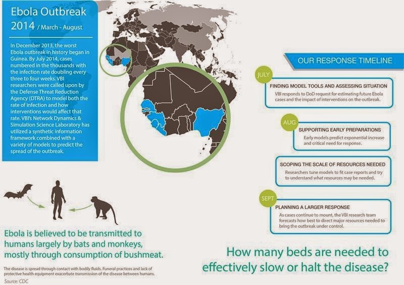
Computational model: Ebola could infect more than 1.4 million people by end of January 2015

Written By Unknown on Wednesday, January 7, 2015 | 7:21 AM

The Network Dynamics and Simulation Science Laboratory at the Virginia Bioinformatics Institute modeled the rate of infections and how interventions would affect the rate.
Credit: Source: CDC / Image courtesy of Virginia Tech
The Ebola epidemic could claim hundreds of thousands of lives and infect more than 1.4 million people by the end of January, according to a statistical forecast released this week by the U.S. Centers for Disease Control and Prevention.

The CDC forecast supports the drastically higher projections released earlier by a group of scientists, including epidemiologists with the Virginia Bioinformatics Institute, who modeled the Ebola spread as part of a National Institutes of Health-sponsored project called Midas, short for Models of Infectious Disease Agent Study.

The effort is also supported by the federal Defense Threat Reduction Agency.
Before the scientists released results, the outbreak in West Africa was expected to be under control in nine months with only about 20,000 total cases. But modeling showed 20,000 people could be infected in just a single month.

The predictions could change dramatically if public health efforts become effective, but based on the virus's current uncontrolled spread, numbers of people infected could skyrocket.

"If the disease keeps spreading as it has been we estimate there could be hundreds of thousands of cases by the end of the year in Liberia alone," said Bryan Lewis, a computational epidemiologist with the Network Dynamics and Simulation Science Laboratory at the Virginia Bioinformatics Institute.

Lewis and his fellow researchers use a combination of models to predict outcomes of the epidemic.

The agent-based models are adaptive, evolving as more information is fed into them to provide an accurate forecast.

Pharmaceutical intervention, which is still on the horizon, is proving less effective in the models than supportive care and personal protection equipment for health care workers.

"The work with Ebola is not an isolated event," said Christopher Barrett, the executive director of the institute. "This research is part of a decades-long effort largely funded by the Defense Threat Reduction Agency to build a global synthetic population that will allow us to ask questions about our world and ourselves that we have never been able to ask before, and to use those answers to prevent or quickly intervene during a crisis."

Barrett and other institute leaders updated U.S. Sen. Tim Kaine and Virginia Tech President Timothy Sands about the Network Dynamics and Simulation Science Lab's role in analyzing the Ebola outbreak at the Virginia Tech Research Center in Arlington on Tuesday morning. That afternoon in Blacksburg they briefed staff members from U.S. Sen. Mark Warner's office.

A university-level Research Institute of Virginia Tech, the Virginia Bioinformatics Institute was established in 2000 with an emphasis on informatics of complex interacting systems scaling the microbiome to the entire globe. It helps solve challenges posed to human health, security, and sustainability. Headquartered at the Blacksburg campus, the institute occupies 154,600 square feet in research facilities, including state-of-the-art core laboratory and high-performance computing facilities, as well as research offices in the Virginia Tech Research Center in Arlington, Virginia.

Source: Virginia Tech

Trial confirms Ebola vaccine candidate safe, equally immunogenic in Africa

Written By Unknown on Sunday, December 28, 2014 | 7:35 PM

"This is the first study to show comparable safety and immune response of an experimental Ebola vaccine in an African population," says lead author Dr Julie Ledgerwood. "This is particularly encouraging because those at greatest risk of Ebola live primarily in Africa, and diminished vaccine protection in African populations has been seen for other diseases." Credit: © nito / Fotolia
Two experimental DNA vaccines to prevent Ebola virus and the closely related Marburg virus are safe, and generated a similar immune response in healthy Ugandan adults as reported in healthy US adults earlier this year. The findings, from the first trial of filovirus vaccines in Africa, are published in The Lancet.

"This is the first study to show comparable safety and immune response of an experimental Ebola vaccine in an African population," says lead author Dr Julie Ledgerwood from the National Institutes of Allergy and Infectious Diseases (NIAID) at the National Institutes of Health, USA. "This is particularly encouraging because those at greatest risk of Ebola live primarily in Africa, and diminished vaccine protection in African populations has been seen for other diseases."

Scientists from the NIAID developed the DNA vaccines that code for Ebola virus proteins from the Zaire and Sudan strains and the Marburg virus protein. The vaccines contain the construction plans for the proteins on the outer surface of the virus. Immune responses against these proteins have shown to be highly protective in non-human primate models.

In this phase 1 trial, the Makerere University Walter Reed Program enrolled 108 healthy adults aged between 18 and 50 from Kampala, Uganda between November, 2009 and April, 2010. Each volunteer was randomly assigned to receive an intramuscular injection of either the Ebola vaccine (30 volunteers), Marburg vaccine (30), both vaccines (30), or placebo (18) at the start of the study, and again 4 weeks and 8 weeks later.

The vaccines given separately and together were safe and stimulated an immune response in the form of neutralising antibodies and T-cells against the virus proteins. Four weeks after the third injection, just over half of the volunteers (57%; 17 of 30) had an antibody response to the Ebola Zaire protein as did 14 of 30 participants who received both the Ebola and Marburg vaccines. 

However, the antibodies were not long-lasting and returned to undetectable levels within 11 months of vaccination.

Both DNA vaccines were well tolerated in Ugandan adults with similar numbers of local and systemic reactions reported in all groups. Only one serious adverse event (neutropenia; low white blood cell count) was reported in a Marburg vaccine only recipient, but was not thought to be vaccine related.

According to Dr Ledgerwood, "These findings have already formed the basis of a more potent vaccine, delivered using a harmless chimpanzee cold virus, which is undergoing trials in the USA, UK, Mali, and Uganda in response to the ongoing Ebola virus outbreak."

Writing in a linked Comment, Dr Saranya Sridhar from the Jenner Institute at the University of Oxford in the UK says, "[This] study deserves to be the focal point around which the broader question of vaccine development, particularly for Africa, must be addressed. With the uncharitable benefit of hindsight in view of the evolving 2014 Ebola outbreak, we must ask ourselves whether a filovirus vaccine should have been in more advanced clinical development. The international response to the present Ebola outbreak is an exemplar of the speed and purpose with which clinical vaccine development can progress and has set the benchmark against which future vaccine development must be judged. This study is the first step on the aspirational road towards the deployment of filovirus vaccines in Africa and must serve to shake the metaphorical cobwebs that can stall our advance towards this destination."

Source: The Lancet

Doctor who survived Ebola received experimental drug treatment

On 28 September, 2014, a 38-year old doctor, who was in charge of an Ebola virus treatment unit in Lakka, Sierra Leone, developed a fever and diarrhea. He tested positive for the virus on the same day. He was placed on a ventilator and on kidney dialysis, and was given antibiotics together with a 3-day course of an experimental drug called FX06 -- a fibrin-derived peptide that has been shown to reduce vascular leakage and its complications in mice with Dengue hemorrhagic shock. Credit: © nito / Fotolia
On 28 September, 2014, the 38-year old doctor, who was in charge of an Ebola virus treatment unit in Lakka, Sierra Leone, developed a fever and diarrhea. He tested positive for the virus on the same day. The doctor was airlifted to Frankfurt University Hospital on the 5th day of his illness and admitted to a specialized isolation unit.

Within 72 hours of admission he developed signs of vascular leakage and severe multi-organ failure, including the lungs, kidneys, and gastrointestinal tract. He was placed on a ventilator and on kidney dialysis, and was given antibiotics together with a 3-day course of an experimental drug called FX06 -- a fibrin-derived peptide that has been shown to reduce vascular leakage and its complications in mice with Dengue hemorrhagic shock.

A marked improvement in vascular and respiratory function was seen under the combined measures of intensive care and drug treatment. After a 30-day observation period, no Ebola virus genetic material was detected in the patient's blood plasma. The patient was released from hospital and is now with his family.
"Even though the patient was critically ill, we were able to support him long enough for his body to start antibody production and for the virus to be cleared by his body's defenses," explains Dr Wolf.

"In terms of improving treatment for Ebola patients, we have shown how intensive care medicine can successfully be applied under strict isolation conditions," adds senior author Professor Zacharowski, head of the Department of Anaesthetics and Intensive Care Medicine at Frankfurt University Hospital."
The authors conclude by calling for FX06 to be evaluated in clinical trials.

Source: The Lancet

How llamas' unusual antibodies might help in the fight against HIV/AIDS

Llamas contribute to the fight against AIDS. Credit: Nika Stropakke, CC-BY
Most vaccines work by inducing an immune response characterized by neutralizing antibodies against the respective pathogen. An effective HIV vaccine has remained elusive so far, but researchers have continued to make progress, often employing innovative methods. A study published on December 18th in PLOS Pathogens reports that a combination of antibodies from llamas can neutralize (destroy) a wide range of circulating HIV viruses.

After initial disappointment that HIV vaccine candidates were unable to elicit neutralizing antibodies, researchers found that some HIV-infected individuals did produce such antibodies. The current challenge is therefore to find safe and effective vaccine formulations (as opposed to HIV infection) that trigger the development of neutralizing antibodies that can recognize and prevent infection with all or most circulating HIV subtypes.

Many known neutralizing antibodies are directed against a specific part of the virus that binds to the CD4 receptor on the human target cells, and structural biology studies indicated that the site is a narrow groove. Antibodies in most mammals are relatively large proteins made up of two copies of two different individual parts (or chains), and bulkiness might be one reason why neutralizing antibodies are rare. Llamas are a notable exception: besides the common four-chain antibodies they also produce smaller ones made up of only two of the four chains. Robin Weiss, an HIV expert, and Theo Verrips, a llama antibody expert, therefore started working with this unconventional research animal.

Laura McCoy (working with Weiss at University College London, UK) led an international group of researchers to test immunization protocols and the resulting immune response in llamas. Having previously identified one particular HIV neutralizing llama antibody, for this study the researchers immunized two additional llamas and identified a total of three new neutralizing antibodies. The four HIV neutralizing llama antibodies target different parts of the CD4-binding site of the virus, and the researchers could show that when used in combination, rather than interfering with each other, they are more potent and can neutralize all of the 60 different HIV strains tested.

To understand how the llama immunization--which included two sets of four sequential vaccine injections per animal--worked, the researchers sequenced many copies of antibody-coding genes from blood cells collected after the first set of immunizations and after a further four rounds of vaccination. They also looked at the "naïve" antibody repertoire from seven llamas that had not been vaccinated. The results suggest that the neutralizing antibodies were not part of the pre-immunization repertoire, nor were they detectable after the first vaccination round. Rather, they were generated as immune cells repeatedly encountered the vaccine and responded by maturing specific antibodies that can recognize it.

While it is encouraging that broadly neutralizing antibodies were found in all of the immunized llamas, they are present only at low concentrations in the blood, and so fail to meet the goal for a protective HIV vaccine. Nonetheless, the researchers conclude that the llama model has allowed them to examine the generation of four broadly neutralizing antibodies induced by vaccination, which has not been possible in any other species.

Source: PLOS.

Genetic factors behind surviving or dying from Ebola shown in mouse study

Written By Unknown on Thursday, December 25, 2014 | 2:06 AM

In an emerging disease research lab at the University of Washington, Chris Williams, a research scientist who specializes in microbiology laboratory robotics, programs a piece of equipment that can be programmed to performs many lab tasks. Credit: Brian Donohue
A newly developed mouse model suggests that genetic factors are behind the mild-to-deadly range of reactions to the Ebola virus.

People exposed to Ebola vary in how the virus affects them. Some completely resist the disease, others suffer moderate to severe illness and recover, while those who are most susceptible succumb to bleeding, organ failure and shock.
In earlier studies of populations of people who have contracted Ebola, these differences are not related to any specific changes in the Ebola virus itself that made it more or less dangerous; instead, the body's attempts to fight infection seems to determine disease severity.

In the Oct. 30 edition of Science, scientists describe strains of laboratory mice bred to test the role of an individual's genetic makeup in the course of Ebola disease. Systems biologists and virologists Angela Rasmussen and Michael Katze from the Katze Laboratory at the University of Washington Department of Microbiology led the study in collaboration with the National Institutes of Health's Rocky Mountain Laboratories in Montana and University of North Carolina at Chapel Hill.

Research on Ebola prevention and treatment has been hindered by the lack of a mouse model that replicates the main characteristics of human Ebola hemorrhagic fever. The researchers had originally obtained this genetically diverse group of inbred laboratory mice to study locations on mouse genomes associated with influenza severity.

The research was conducted in a highly secure, state-of-the-art biocontainment safety level 4 laboratory in Hamilton, Mont. The scientists examined mice that they infected with a mouse form of the same species of Ebola virus causing the 2014 West Africa outbreak. The study was done in full compliance with federal, state, and local safety and biosecurity regulations. This type of virus has been used several times before in research studies. Nothing was done to change the virus.

Interestingly, conventional laboratory mice previously infected with this virus died, but did not develop symptoms of Ebola hemorrhagic fever.

In the present study, all the mice lost weight in the first few days after infection. Nineteen percent of the mice were unfazed. They not only survived, but also fully regained their lost weight within two weeks. They had no gross pathological evidence of disease. Their livers looked normal.

Eleven percent were partially resistant and less than half of these died. Seventy percent of the mice had a greater than 50 percent mortality. Nineteen percent of this last group had liver inflammation without classic symptoms of Ebola, and thirty-four percent had blood that took too long to clot, a hallmark of fatal Ebola hemorrhagic fever in humans. Those mice also had internal bleeding, swollen spleens and changes in liver color and texture.
The scientists correlated disease outcomes and variations in mortality rates to specific genetic lines of mice.

"The frequency of different manifestations of the disease across the lines of these mice screened so far are similar in variety and proportion to the spectrum of clinical disease observed in the 2014 West African outbreak," Rasmussen said.

While acknowledging that recent Ebola survivors may have had immunity to this or a related virus that saved them during this epidemic, Katze said, "Our data suggest that genetic factors play a significant role in disease outcome."

In general, when virus infection frenzied the genes involved in promoting blood vessel inflammation and cell death, serious or fatal disease followed. On the other hand, survivors experienced more activity in genes that order blood vessel repair and the production of infection-fighting white blood cells.

The scientists note that certain specialized types of cells in the liver could also have limited virus reproduction and put a damper on systemic inflammation and blood clotting problems in resistant mice. Susceptible mice had widespread liver infection, which may explain why they had more virus in their bodies and poorly regulated blood coagulation. The researchers also noticed that spleens in the resistant and susceptible mice took alternate routes to try to ward off infection.

"We hope that medical researchers will be able to rapidly apply these findings to candidate therapeutics and vaccines," Katze said. They believe this mouse model can be promptly implemented to find genetic markers, conduct meticulous studies on how symptoms originate and take hold, and evaluate drugs and that have broad spectrum anti-viral activities against all Zaire ebolaviruses, including the one responsible for the current West African epidemic.

Experimental Ebola vaccine appears safe, prompts immune response

Written By Unknown on Wednesday, December 24, 2014 | 12:56 AM

A 39-year-old woman, the first participant enrolled in VRC 207, receives a dose of the investigational NIAID/GSK Ebola vaccine at the NIH Clinical Center in Bethesda, Md. on September 2. Credit: NIAID
An experimental vaccine to prevent Ebola virus disease was well-tolerated and produced immune system responses in all 20 healthy adults who received it in a Phase 1 clinical trial conducted by researchers from the National Institutes of Health. The candidate vaccine, which was co-developed by the NIH's National Institute of Allergy and Infectious Diseases (NIAID) and GlaxoSmithKline (GSK), was tested at the NIH Clinical Center in Bethesda, Maryland. The interim results are reported online in advance of print in the New England Journal of Medicine.

"The unprecedented scale of the current Ebola outbreak in West Africa has intensified efforts to develop safe and effective vaccines, which may play a role in bringing this epidemic to an end and undoubtedly will be critically important in preventing future large outbreaks," said NIAID Director Anthony S. Fauci, M.D. "Based on these positive results from the first human trial of this candidate vaccine, we are continuing our accelerated plan for larger trials to determine if the vaccine is efficacious in preventing Ebola infection."

The candidate NIAID/GSK Ebola vaccine was developed collaboratively by scientists at the NIAID Vaccine Research Center (VRC) and at Okairos, a biotechnology company acquired by GSK. It contains segments of Ebola virus genetic material from two virus species, Sudan and Zaire. The Ebola virus genetic material is delivered by a carrier virus (chimpanzee-derived adenovirus 3 or cAd 3) that causes a common cold in chimpanzees but causes no illness in humans. The candidate vaccine does not contain Ebola virus and cannot cause Ebola virus disease.

The trial enrolled volunteers between the ages of 18 and 50. Ten volunteers received an intramuscular injection of vaccine at a lower dose and 10 received the same vaccine at a higher dose. At two weeks and four weeks following vaccination, the researchers tested the volunteers' blood to determine if anti-Ebola antibodies were generated. All 20 volunteers developed such antibodies within four weeks of receiving the vaccine. Antibody levels were higher in those who received the higher dose vaccine.

The investigators also analyzed the research participants' blood to learn whether the vaccine prompted production of immune system cells called T cells. A recent study by VRC scientist Nancy J. Sullivan, Ph.D., and colleagues showed that non-human primates inoculated with the candidate NIAID/GSK vaccine developed both antibody and T-cell responses, and that these were sufficient to protect vaccinated animals from disease when they were later exposed to high levels of Ebola virus.

The experimental NIAID/GSK vaccine did induce a T-cell response in many of the volunteers, including production of CD8 T cells, which may be an important part of immune protection against Ebola viruses. Four weeks after vaccination, CD8 T cells were detected in two volunteers who had received the lower dose vaccine and in seven of those who had received the higher dose.

"We know from previous studies in non-human primates that CD8 T cells played a crucial role in protecting animals that had been vaccinated with this NIAID/GSK vaccine and then exposed to otherwise lethal amounts of Ebola virus," said Julie E. Ledgerwood, D.O., a VRC researcher and the trial's principal investigator. "The size and quality of the CD8 T cell response we saw in this trial are similar to that observed in non-human primates vaccinated with the candidate vaccine."

There were no serious adverse effects observed in any of the volunteers, although two people who received the higher dose vaccine did develop a briefly lasting fever within a day of vaccination.

Using power of computers to harness human genome may provide clues into Ebola virus

Written By Unknown on Tuesday, December 23, 2014 | 5:39 PM

Ramaswamy Narayanan, Ph.D., professor in the Charles E. Schmidt College of Science at Florida Atlantic University.
Ramaswamy Narayanan, Ph.D., professor in the Charles E. Schmidt College of Science at Florida Atlantic University, is working to blend the power of computers with biology to use the human genome to remove much of the guesswork involved in discovering cures for diseases.

In an article titled "Ebola-Associated Genes in the Human Genome: Implications for Novel Targets," published in the current MedCrave Online Journal of Proteomics and Bioinformatics, Narayanan describes how key genes that are present in our cells could be used to develop drugs for this disease.

"Bioinformatics is a powerful tool to help us understand biological data," said Narayanan whose research has focused in this field for more than a decade. "We are mining the human genome for Ebola virus association to develop an understanding of the human proteins involved in this disease for subsequent research and development, and to potentially create a pipeline of targets that we can test and evaluate."

Ebola virus disease is a major healthcare challenge facing the globe today and if left unchecked could become a pandemic. A limited knowledgebase exists about the Ebola virus and companies are hastening to develop vaccines and other forms to treat and cure the virus. There are no FDA-approved drugs, and developing vaccines or antibodies and testing them in clinical trials is an arduous process that takes considerable time. Currently, patients infected with Ebola are only able to receive supportive care such as fluid replacement, nutritional support, pain control, and blood pressure maintenance. In some cases, patients may be fortunate enough to be treated with experimental drugs.

Narayanan's work has helped to identify numerous FDA-approved drugs already used for many other diseases including anti-inflammatory drugs, anticoagulants, cancer, HIV, statins and hormones, which could potentially be used to add to the current supportive care for patients with the Ebola virus.

"With the high mortality rate of this disease, the world urgently needs new ways to treat patients," said Narayanan. "The ability to use drugs that are already approved by the FDA could provide clinicians with more options to treat Ebola patients, rather than just relying on supportive measures like fluid replacement or antibiotics."

According to the World Health Organization (WHO), Ebola virus disease (EVD) is a severe, often fatal illness in humans. The virus is transmitted to people from wild animals and spreads in the human population through human-to-human transmissions. The evolving knowledge of this disease is prompting appropriate attention locally and globally. The 2014 Ebola epidemic has affected multiple countries in West Africa with some cases observed in Europe and the United States.

Source: Florida Atlantic University

Researchers ferret out a flu clue

Written By Unknown on Monday, December 22, 2014 | 9:10 PM

Professor Michael Jennings, Deputy Director of the Institute for Glycomics. Credit: Image courtesy of Griffith University
Research that provides a new understanding as to why ferrets are similar to humans is set to have major implications for the development of novel drugs and treatment strategies.

Published in the journal Nature Communications, the research is a collaboration between Professor Michael Jennings and other researchers from the Institute for Glycomics, Griffith University and collaborators at the University of Queensland and the University of Adelaide.

The team has shown for the first time that ferrets share a mutation that was previously thought to be unique to humans, among the mammals. This helps to explain why the molecular characteristics of ferrets so uniquely mimic human susceptibility, severity and transmission of influenza A virus strains.

Professor Michael Jennings, Deputy Director of the Institute for Glycomics, says these findings open up a completely novel approach to tackling human diseases from influenza through to cancer.

"For over 80 years we've known that ferrets are uniquely susceptible to human influenza A virus, but the precise reason was unknown," Professor Jennings said.

"We have shown that ferrets have a mutation in a gene required to make a crucial sugar called sialic acid. Most animals can make two types of sialic acid. Ferrets, like humans can make only one. Different flu strains have preferences for the type of sialic acid they bind to cause infection. Because ferrets can only make the human form of this sugar, they are naturally "humanized" for the receptors recognised by human strains of the flu virus."

Source: Griffith University

Flu virus key machine: First complete view of structure revealed

The complete structure allows researchers to understand how the polymerase uses host cell RNA (red) to kick-start the production of viral messenger RNA. Credit: © EMBL/P.Riedinger
Scientists looking to understand -- and potentially thwart -- the influenza virus now have a much more encompassing view, thanks to the first complete structure of one of the flu virus' key machines. The structure, obtained by scientists at EMBL Grenoble, allows researchers to finally understand how the machine works as a whole, and could prove instrumental in designing new drugs to treat serious flu infections and combat flu pandemics.

If you planned to sabotage a factory, a recon trip through the premises would probably be much more useful than just peeping in at the windows. Scientists looking to understand -- and potentially thwart -- the influenza virus have now gone from a similar window-based view to the full factory tour, thanks to the first complete structure of one of the flu virus' key machines. The structure, obtained by scientists at the European Molecular Biology Laboratory (EMBL) in Grenoble, France, allows researchers to finally understand how the machine works as a whole. Published in two papers in Nature, the work could prove instrumental in designing new drugs to treat serious flu infections and combat flu pandemics.

The machine in question, the influenza virus polymerase, carries out two vital tasks for the virus. It makes copies of the virus' genetic material -- the viral RNA -- to package into new viruses that can infect other cells; and it reads out the instructions in that genetic material to make viral messenger RNA, which directs the infected cell to produce the proteins the virus needs. Scientists -- including Cusack and collaborators -- had been able to determine the structure of several parts of the polymerase in the past. But how those parts came together to function as a whole, and how viral RNA being fed in to the polymerase could be treated in two different ways remained a mystery.

"The flu polymerase was discovered 40 years ago, so there are hundreds of papers out there trying to fathom how it works. But only now that we have the complete structure can we really begin to understand it," says Stephen Cusack, head of EMBL Grenoble, who led the work.

Using X-ray crystallography, performed at the European Synchrotron Radiation Facility (ESRF) in Grenoble, Cusack and colleagues were able to determine the atomic structure of the whole polymerase from two strains of influenza: influenza B, one of the strains that cause seasonal flu in humans, but which evolves slowly and therefore isn't considered a pandemic threat; and the strain of influenza A -- the fast-evolving strain that affects humans, birds and other animals and can cause pandemics -- that infects bats.

"The high-intensity X-ray beamlines at the ESRF, equipped with state-of-the-art Dectris detectors, were crucial for getting high quality crystallographic data from the weakly diffracting and radiation sensitive crystals of the large polymerase complex," says Cusack. "We couldn't have got the data at such a good resolution without them."

The structures reveal how the polymerase specifically recognises and binds to the viral RNA, rather than just any available RNA, and how that binding activates the machine. They also show that the three component proteins that make up the polymerase are very intertwined, which explains why it has been very difficult to piece together how this machine works based on structures of individual parts.

Although the structures of both viruses' polymerases were very similar, the scientists found one key difference, which showed that one part of the machine can swivel around to a large degree. That ability to swivel explains exactly how the polymerase uses host cell RNA to kick-start the production of viral proteins. The swivelling component takes the necessary piece of host cell RNA and directs it into a slot leading to the machine's heart, where it triggers the production of viral messenger RNA.

Now that they know exactly where each atom fits in this key viral machine, researchers aiming to design drugs to stop influenza in its tracks have a much wider range of potential targets at their disposal -- like would-be saboteurs who gain access to the whole production plant instead of just sneaking looks through the windows. And because this is such a fundamental piece of the viral machinery, not only are the versions in the different influenza strains very similar to each other, but they also hold many similarities to their counterparts in related viruses such as lassa, hanta, rabies or ebola.

The EMBL scientists aim to explore the new insights this structure provides for drug design, as well as continuing to try to determine the structure of the human version of influenza A, because although the bat version is close enough that it already provides remarkable insights, ultimately fine-tuning drugs for treating people would benefit from/require knowledge of the version of the virus that infects humans. And, since this viral machine has to be flexible and change shape to carry out its different tasks, Cusack and colleagues also want to get further snapshots of the polymerase in different states.

"This doesn't mean we now have all the answers," says Cusack, "In fact, we have as many new questions as answers, but at least now we have a solid basis on which to probe further."

Source: European Molecular Biology Laboratory (EMBL)
 
Support : Creating Website | Johny Template | Mas Template
Copyright © 2011. The planet wall - All Rights Reserved
Template Created by Easy Blogging Published by Mas Template
Proudly powered by Blogger