Latest Post
Showing posts with label INFECTIOUS DISEASES. Show all posts

HIV virus in disguise tricks immune system, Marie Larsson is Professor of Molecular Virology

Written By Unknown on Friday, February 6, 2015 | 3:17 PM

Marie Larsson is Professor of Molecular VirologyName: Marie LarssonTitle: Professor of Molecular Virology
Department: IKE

CONTACT

Phone: +46 (0)10-103 10 55
E-mail: marie.larsson@liu.se
Address:
Linköping University
Department of Clinical and Experimental Medicine
Virology
SE-581 85 Linköping
Sweden

Marie Larsson is Professor of Molecular Virology. Her research is in the area of immunovirology, specifically HIV research with focus on the immunomodulatory effect this virus has on dendritic cells and T cells. She has also ongoing projects exploring new adjuvants and vaccine constellations for cancer and virus. Furthermore, she is investigating the induction and sustainment of cancer associated inflammation and the deleterious effect this has on host immune defense.

Immunomodulatory effects of HIV-1’s interactions with DCs and T cells from
HIV virus

blood and mucosa
So far over 30 million people have died from HIV-1 infection (figure 1), the majority of them in the developing countries, and this epidemic is still cause for major concern. The existing antiretroviral therapy dampens the infection and the destruction of the immune system, i.e. AIDS, but does not cure the disease. Sadly, this therapy is not available to all HIV infected and is a very expensive lifelong commitment with severe side effects.

HIV virusA vaccine blocking HIV infection is theDendritic cell sought-after solution but there is no hope that we will have such a vaccine in the near future. Instead we can hope for a therapy that induces a potent long lasting immune response consisting of CD4+ and CD8+ T cells, two types of control cells involved in the immune defense, that have proven to be important to control the infection. There exists a unique cell in all tissues in our bodies, the dendritic cell (DC) (Figure 2) with unique ability to activate T cells so they can perform their job in the body. DCs in the vaginal and rectal tissues are one of the first cells to encounter HIV during intercourse with an infected individual (Figure 3 and 4). Unfortunately, HIV hijacks the DCs, which makes this cell responsible for spreading the virus to interacting T cells in the body which provokes HIV-infection of T cells and cell death when it should be initiating immune responses to fight the infection.DC HIV

HIV virusMy research aspires to elucidate the mechanisms behind the immunomodulatory effects HIV exerts on DCs and on their ability to activate T cells. Focus will be on; Elucidation of the mechanisms involved in HIV’s binding to and uptake by DCs and the subsequent degradation that leads to DC antigen presentation of HIV peptides for activation of HIV specific T cells. Elucidation of the mechanisms responsible for the negative effects HIV exerts on DCs and if presence of HIV virions during DC T cell priming impairs the T cell function. Elucidate the effect opsonized HIV-1 exerts on immune cells such as DCs, NK cells and T cells. Identification of receptors and cells involved in the initial HIV infection of cervical mucosa and colorectal mucosa,  and potential microbiocides that can block the initial infection, and elucidation of why HIV affects the T cells in the gut to a higher extent than the T cells in blood.

HIV will continue to kill people and have a great impact on mankind until we have a drug that can stop this infection. My ambition is that the planed research will answer some basic questions regarding the role of DCs in HIV pathogenesis and induction of potent immune response against this virus. This knowledge will guide how a vaccine/therapy needs to be constructed in order to have high efficacy.
Mucosal transmission
                                                             Mucosal transmission
                                                                Cervix

Cancer research

Elucidation the role of IL-1α and the microenvironment in development of pancreatic cancer

Pancreatic ductal adenocarcinoma (PDAC) is a common gastrointestinal malignancy with an exceptional poor prognosis and a mortality rate that nearly matches the rate of incidence. The cross-talk between PDAC and stroma cells, e.g. cancer associated fibroblasts (CAF), and immune cells, may create an environment with chronic inflammation augmenting tumor transformation and maintenance.

In PDAC, more than 70% of the total tumor mass can consist of fibrotic stroma, which makes CAFs the major component in this cancer. PDAC inflammatory environment consists of many mediators, e.g. IL-1, COX-2, IL-6, and CXCL8, and some of these factors correlate to tumor development and poor prognosis. Of note, elevated expression of IL-1 in tumors has been associated with more aggressive disease. Several studies, including ours have reported that dendritic cells (DCs), one of the immune cells found in the tumor microenvironment, show phenotypic and functional abnormalities when isolated from tumor bearing animals and individuals with PDAC. Recent findings provided some evidence that the COX-2 metabolite PGE2 is involved in the upregulation of immunomodulatory factors in DCs impairing their T cell stimulatory ability. The aims are to examine the receptor-ligands and signaling pathways involved in the cross talk between stroma cells, i.e. CAFs, and PDAC cells giving rise to the inflammatory environment and creating an environment sustaining the PDAC. To examine the role IL-1 cytokine family and effects these cytokines cell signaling have on creating the inflammatory environment and in tumor development and survival. To examine whether neutralization of IL1 signaling pathway enhances the survival and quality of life for individuals with pancreatic cancer.

The information gained from the proposed research will help us understand the mechanisms underlying the development of PDAC and the effect this solid tumor exert on the body and may help designing therapies for PDAC.

Source: Linköping University

Scheduling tool a big help in healthcare

The HIV virus avoids the body’s immune cells by disguising itself using proteins that normally take part in the defence against infections. These are the findings of research conducted at the Division of Molecular Virology.
Professor Marie Larsson

Professor Marie LarssonThese “complement proteins” are soluble molecules that attach themselves to foreign particles and those of the body in different patterns. These patterns help the immune system to identify and attack dangerous intruders such as viruses and bacteria.

Phd student Rada EllegårdInstead, HIV exploits the complement proteins present in all body fluids in order to make its way into the body tissues without being attacked. It was known previously that infections are much more effective if the virus is surrounded by the bodily fluids normally present in blood or sexual contagion than if it is isolated in a laboratory environment. In an article in the Journal of Immunology Marie Larsson, professor of molecular virology (left) and PhD student Rada Ellegård (right) provide an explanation of this phenomenon.

“The ability to use our complement proteins in this way is probably key to the success of HIV in being transmitted. We are currently carrying out further work to investigate these mechanisms in the male genital mucous membrane – the tissue where the virus infection takes place in sexual transmission,” says Ms Larsson.
rada


The first thing HIV particles come into contact with is dendritic cells, immune cells that function as sentry posts. Their job is to identify viruses as dangerous and hostile to the body, and to respond by producing the substances that combat infections and moving to a lymph node to set a specific immune defence in motion.

In the case of HIV, this process does not seem to work very well. The disguise stops the dendritic cells recognising the virus, which instead establishes an infection in the mucous membrane and uses the movement to the lymph nodes as a way to spread around the whole body.

As more and more cells are infected and killed, the virus slowly breaks down our immune system. Without treatment this sequence of events leads to AIDS, a condition where the victim becomes extremely vulnerable to infection and where normally harmless viruses and bacteria become life-threatening.

Related content

Marie Larsson - research presentation >>

HIV-illustration
                                       HIV-illustration

When a virus is captured by a dendritic cell, it is recognised by a virus sensor that normally sets in motion infection reduction factors which inhibit the production of new virus particles (picture, left). HIV has the ability to clothe itself in complements, a type of protein present in our bodily fluids. In this way it can dampen the signals from the virus sensors and produce many new particles (picture, right).


Source: Linköping University
New Molecular Target Identified for Treating Cerebral Malaria

Written By Unknown on Thursday, February 5, 2015 | 5:16 AM

Mosquito Anopheles
                                                             Mosquito Anopheles

A drug already approved for treating other diseases may be useful as a treatment for cerebral malaria, according to researchers at Harvard T. H. Chan School of Public Health. They discovered a novel link between food intake during the early stages of infection and the outcome of the disease, identifying two molecular pathways that could serve as new targets for treatment.

“We have known for a long time that nutrition can affect the course of infectious disease, but we were surprised at how rapidly a mild reduction in food intake could improve outcome in a mouse malaria model,” said senior author James Mitchell, associate professor of genetics and complex diseases. “However, the real importance of this work is the identification of unexpected molecular pathways underlying cerebral malaria that we can now target with existing drugs.”

The study appears online January 30, 2015 in Nature Communications.

Cerebral malaria — a severe form of the disease — is the most serious consequence of infection by the parasite Plasmodium falciparum, resulting in seizures, coma, and death. Currently there is a lack of safe treatment options for cerebral malaria, particularly for use in children, who represent the majority of cases. Even patients who receive early treatment with standard antimalarial chemotherapeutic agents run a high risk of dying, despite clearance of the parasite. Moreover, around 25% of survivors develop neurological complications and cognitive impairment.

Lead authors Pedro Mejia and J. Humberto Treviño-Villarreal, both researchers at Harvard T.H. Chan School of Public Health, found that leptin—a hormone secreted from fat tissue with roles in suppressing appetite, but also in activating adaptive immune and inflammatory responses—is increased upon infection in a mouse model of cerebral malaria, and turns out to be a major bad actor in promoting neurological symptoms and death. Remarkably, Mejia, Treviño-Villarreal and colleagues showed that reducing leptin using a variety of means, either genetically, pharmacologically, or nutritionally by reducing food intake during the first two days of infection, protected against cerebral malaria.

The researchers also found that leptin acted primarily on cytotoxic T cells by turning on the well-studied mTOR protein, for which pharmacologic inhibitors are readily available. In their animal model, treating mice with the mTOR inhibitor rapamycin protected them against the neurological complications of cerebral malaria. Protection was due in part to a preservation of the blood brain barrier, which prevented the entry of blood cells carrying the parasites into the brain. As rapamycin is already FDA-approved for use in humans, trials in humans for cerebral malaria treatment with this drug may be possible, according to the researchers.

This study was the result of an ongoing collaboration between the Mitchell lab in the Department of Genetics and Complex Diseases and the labs of Manoj Duraisingh and Dyann Wirth in the Department of Immunology and Infectious Diseases. Other Harvard T.H. Chan School of Public Health authors included  Christopher Hine, Eylul Harputlugil, Samantha Lang, Ediz Calay and Rick Rogers.

This study was supported in part by grants from NIH (DK090629 and AG036712) and the Glenn Foundation for Medical Research to J.R.M.; a Harvard T.H. Chan School of Public Health Yerby postdoctoral fellowship to Mejia, and financial support from the Universidad Auto´noma de Nuevo Leo´n to Treviño-Villarreal.

Source: Harvard

Ebola: Reports from the front lines

Written By Unknown on Friday, January 30, 2015 | 4:21 AM

Dr. Noah Rosenberg “Ebola was never the only killer here, and our ability to appropriately diagnose and treat these patients is woefully limited.”
Alpert Medical School professors Michael Smit and Noah Rosenberg are in Sierra Leone and Liberia respectively, treating Ebola patients. There are some signs of a slowdown in the epidemic, but the doctors emphasize that the virus must be fought “until the last case.”

PROVIDENCE, R.I. [Brown University] — In a limited sense, two Brown University medical professors who have been fighting Ebola in West Africa this winter have good news to report. They have seen some signs of slowing disease transmission. But the broader reality of what Ebola has done in Sierra Leone and Liberia is grim, according to Drs. Michael Smit and Noah Rosenberg.

Ebola shouldn’t just be contained, they note: It must be treated until all cases are resolved.

Smit, assistant professor of pediatrics and a physician at Hasbro Children’s Hospital, arrived in Sierra Leone Dec. 3, 2014, and will remain there through Jan. 18. Rosenberg, clinical assistant professor of emergency medicine and a Lifespan doctor, arrived in Liberia in mid-December and will stay until Jan. 27. They answered questions for medical science writer David Orenstein about what they are doing and seeing, and what people back in the States need to know.

Please describe a typical day.

NR: I arrive at 7 a.m. and meet with the overnight doctor and nurses to discuss our patients. My team pulls on full personal protective equipment and enters the high-risk area. We examine each patient, inquire about their current symptoms, give routine supportive medications and IV fluids if needed. We take blood to test for Ebola. We remove our equipment while being intermittently sprayed with chlorine. Rounds repeat in the afternoon and we often make an additional trip for a new admission. In the evening we meet with the night team and then rest to return in the morning.
Dr. Michael Smit “The effect of the epidemic goes far beyond those infected with Ebola virus. ... The economic impact on the country is devastating.
MS: I am the medical team leader of one of four medical teams at the Mateneh Ebola Treatment Center (ETC). Our days differ depending on which shift we are working. When working the early shift, we eat breakfast around 7 a.m. and drive to the ETC. We change into our work uniforms of scrubs and rubber boots at the ETC. At 7:30 we receive signout on the patients from the overnight team. We then assign personnel to conduct the nursing rounds, physician rounds, admissions, and discharges. During the heat of the day, we try to limit time in the high-risk area in personal protective equipment (PPE) to one hour. We deliver meals, administer medications, place intravenous catheters, and draw blood for laboratory tests. We triage admissions as they come through the ambulance bay. We also process discharges. These include survivors discharged home and deaths transferred to the morgue. At the end of the shift, we sign out the patients to the oncoming team, change back into our civilian clothes, and go back to the compound to eat. The late and overnight shifts are the same as the early shift for the most part.

What do you observe and hear about the status of the epidemic where you are?

NR: The exponential growth of the epidemic is clearly over in Liberia, but sporadic outbreaks are likely to continue until every case has been eliminated from West Africa, which may take months. Most of our new admissions now test negative for Ebola; they frequently die nonetheless. This may seem counterintuitive, but Ebola was never the only killer here, and our ability to appropriately diagnose and treat these patients is woefully limited.

MS: It is difficult to assess the status of the epidemic from here. We have limited access to the Internet, so our updates of what is happening are intermittent. As for what we observe, after a slow start from our opening in mid-December, we saw a steady increase in cases in our district in Bombali. Over the last week, admissions have decreased. The reason for this is not clear. We hope that it reflects a reduction in transmission, but we cannot assume this given the complexities of case identification and transport here.

What do people in the United States most need to know, based on what you are experiencing?

NR: Before the epidemic Liberia suffered from a large disease burden and severely limited health care system. Decades of civil war, sparked by inequality and tension between descendants of indigenous West Africans and freed slaves, wrecked the infrastructure and economy. Not only was the U.S.A. responsible for the founding of Liberia, but many of the struggles here today have their origins in the slave trade. Ebola will soon fade from the news but the epidemic has only worsened an already grave condition. We have a special responsibility to Liberia and it deserves our sustained attention.

MS: People in the United States need to know that the epidemic here is far from over. Even in a situation with diminished transmission, the United States needs to send personnel and resources here to combat Ebola until the last case. The effect of the epidemic goes far beyond those infected with Ebola virus. The schools here in Sierra Leone have been closed for months. Teen pregnancy is increasing as a result. The economic impact on the country is devastating, with some estimates setting the economy back 10 years. Also, people in the United States need to know about the dedication and resilience of the local and international healthcare workers who are fighting the epidemic, often under challenging physical and emotional conditions.

Source: Brown University

In mice, vaccine stops urinary tract infections linked to catheters

Written By Unknown on Sunday, January 18, 2015 | 3:45 AM

To adhere to catheters and start urinary tract infections, bacteria extend microscopic fibers with sticky proteins at their ends. Scientists have developed a vaccine that blocks the EbpA protein, visible as a white bulge above, and stops infections in mice. Credit: John Heuser
The most common type of hospital-associated infection may be preventable with a vaccine, new research in mice suggests.

The experimental vaccine, developed by researchers at Washington University School of Medicine in St. Louis, prevented urinary tract infections associated with catheters, the tubes used in hospitals and other care facilities to drain urine from a patient's bladder.

Each day a catheter is present in the urethra and the bladder, the risk of urinary tract infection increases. Nearly every patient who has a catheter for more than 30 days acquires a urinary tract infection. The infections make urination painful and can damage the bladder. If untreated, bacteria can cross into the bloodstream and cause sepsis, a potentially life-threatening complication.

"Catheter-associated urinary tract infections are very common," said first author Ana Lidia Flores-Mireles, PhD, a postdoctoral research associate at the School of Medicine. "Antibiotic resistance is increasing rapidly in the bacteria that cause these infections, so developing new treatments is a priority."

The study is available online Sept. 17 in Science Translational Medicine.

Manufacturers typically coat catheters with antibiotics to reduce the risk of infection. But Flores-Mireles and her colleagues in the laboratory of Scott Hultgren, PhD, showed that inserting catheters into the bladder provokes an inflammatory response that results in the catheter being covered with fibrinogen, a blood-clotting protein.

Fibrinogen shields bacteria from the antibiotics and provides bacteria with a landing pad to adhere to and food to consume as they establish an infection, the research revealed.

"The bacteria use long, thin hairs known as pili to anchor themselves to the fibrinogen, and then they can start to form biofilms, which are slimy coatings on the surface of the catheter composed of many bacteria," said co-author Michael Caparon Jr., PhD, professor of molecular microbiology. "The biofilms protect the bacteria from antibiotics and immune cells, further prevent them from being washed from the body by the flow of urine, and make it possible for bacteria to seed the lining of the bladder with infections."

The urethra and bladder of a mouse are too small to insert a full catheter into, but the scientists showed that surgically implanting a small segment of catheter into the bladder via the urethra increased vulnerability to infection in a similar fashion.

Working with Enterococcus faecalis, a common cause of catheter-associated urinary tract infections, Flores-Mireles showed that a protein on the end of the pili, EbpA, binds to fibrinogen and makes it possible for the bacteria to begin forming biofilms.
When Flores-Mireles prevented the bacteria from making EbpA, they couldn't start infections.

"This protein is like the anchor of a boat," she said. "Without the anchor, the infection is at the mercy of the waves and gets washed away."

Next, the researchers injected the mice with a vaccine containing EbpA. The vaccine caused the animal's immune systems to produce antibodies that blocked EbpA and stopped the infectious process.

The scientists are testing to see if the vaccine helps mice clear established infections of E. faecalis. They also are working to develop a monoclonal antibody that blocks EbpA to prevent catheter-associated infections in the urinary tract and elsewhere in the body.

"We took a closer look at this protein and found that one-half of it is essential for binding to fibrinogen to induce infections," Flores-Mireles said. "The segment of genetic code that makes this part of the protein is also found in the genes of many other bacteria that cause urinary tract infections, so a vaccine, antibody or drug that blocks this part of the protein may help prevent other infections linked to catheters in the urinary tract and in other parts of the body."

Cold virus replicates better at cooler temperatures

Written By Unknown on Friday, January 16, 2015 | 8:18 AM

Artist's rendering of a rhinovirus (stock illustration). Credit: © fotoliaxrender / Fotolia
The common cold virus can reproduce itself more efficiently in the cooler temperatures found inside the nose than at core body temperature, according to a new Yale-led study. This finding may confirm the popular yet contested notion that people are more likely to catch a cold in cool-weather conditions.

Researchers have long known that the most frequent cause of the common cold, the rhinovirus, replicates more readily in the slightly cooler environment of the nasal cavity than in the warmer lungs. However, the focus of prior studies has been on how body temperature influenced the virus as opposed to the immune system, said study senior author and Yale professor of immunobiology Akiko Iwasaki.

To investigate the relationship between temperature and immune response, Iwasaki and an interdisciplinary team of Yale researchers spearheaded by Ellen Foxman, a postdoctoral fellow in Iwasaki's lab, examined the cells taken from the airways of mice. They compared the immune response to rhinovirus when cells were incubated at 37 degrees Celsius, or core body temperature, and at the cooler 33 degrees Celsius. "We found that the innate immune response to the rhinovirus is impaired at the lower body temperature compared to the core body temperature," Iwasaki said.

The study also strongly suggested that the varying temperatures influenced the immune response rather than the virus itself. Researchers observed viral replication in airway cells from mice with genetic deficiencies in the immune system sensors that detect virus and in the antiviral response. They found that with these immune deficiencies, the virus was able to replicate at the higher temperature. "That proves it's not just virus intrinsic, but it's the host's response that's the major contributor," Iwasaki explained.

Although the research was conducted on mouse cells, it offers clues that may benefit people, including the roughly 20% of us who harbor rhinovirus in our noses at any given time. "In general, the lower the temperature, it seems the lower the innate immune response to viruses," noted Iwasaki. In other words, the research may give credence to the old wives' tale that people should keep warm, and even cover their noses, to avoid catching colds.

Yale researchers also hope to apply this insight into how temperature affects immune response to other conditions, such as childhood asthma. While the common cold is no more than a nuisance for many people, it can cause severe breathing problems for children with asthma, noted Foxman. Future research may probe the immune response to rhinovirus-induced asthma.

The study was published in the Proceedings of the National Academy of Sciences.

Long-acting drug effectively prevents HIV-like infection in monkeys

The new drug cabotegravir (in vials above) has been shown to protect monkeys from infection by an HIV-like virus, and a clinical trial testing cabotegravir's safety and acceptability has begun. Unlike other preventive treatments, it would require only one injection every three months.
Credit: Zach Veilleux / The Rockefeller University
A regime of anti-HIV drugs -- components of regimens to treat established HIV infection -- has the potential to protect against infection in the first place. But real life can interfere; the effectiveness of this prophylactic approach declines if the medications aren't taken as prescribed.

HIV researchers hope a new compound, known as cabotegravir, could make dosing easier for some because the drug would be administered by injection once every three months. A clinical trial testing long-acting cabotegravir's safety and acceptability has already begun at multiple U.S. sites including The Rockefeller University Hospital. Meanwhile two new studies, including one conducted by researchers at the Aaron Diamond AIDS Research Center (ADARC) and Rockefeller University, published today (January 15) in Science Translational Medicine, show that long-acting cabotegravir injections are highly protective in a monkey model of vaginal transmission of a virus similar to HIV.

"Clinical trial results have demonstrated that the effectiveness of preventive oral medications can range with results as high as 75 percent effective to as low as ineffective, and a lot of that variability appears to hinge on the patient's ability to take the pills as prescribed," says study researcher Martin Markowitz, a professor at Rockefeller University and ADARC. "Long acting cabotegravir has the potential to create an option that could improve adherence by making it possible to receive the drug by injection once every three months."

Developed by ViiV Healthcare and GlaxoSmithKline, and previously known as GSK744 LA, cabotegravir is an antiretroviral drug. Antiretrovirals interfere with HIV's ability to replicate itself using a host cell and they are used to treat an HIV infection or to prevent those at high risk from acquiring it in the first place.

Cabotegravir belongs to a group of antiretrovirals that target integrase, an enzyme the virus uses to integrate itself into the cell's genome. This compound is a relative of an already FDA-approved integrase inhibitor, dolutegravir, but with chemical properties that allow it to be formulated into a long-acting suspension for injection.

A previous study by the ADARC and Rockefeller team in collaboration with ViiV Healthcare and GSK found long-acting cabotegravir could protect male rhesus macaque monkeys from exposure to a virus related to HIV. Following up on these results, a phase 2 clinical trial is now underway in a group of 120 men at low risk of infection. Before cabotegravir's effectiveness in high risk individuals can be tested, trials must show that study participants tolerate the drug well and find the quarterly injections, which are a novel approach to HIV prevention, acceptable.

Both new animal studies were conducted with women in mind; in 2013 women accounted for 47 percent of new HIV infections worldwide according to the Joint United Nations Programme on HIV and AIDS. Working separately, two teams tested the drug's ability to block vaginal transmission in two species of monkeys with different breeding cycles and susceptibility to infection.

First author Chasity Andrews, a postdoctoral fellow at ADARC and Rockefeller, and colleagues at ADARC, the Tulane Regional Primate Center and ViiV/GSK, studied female rhesus macaques treated with progesterone to increase their susceptibility to the virus. They found injections of long acting cabotegravir were 90 percent effective at protecting the monkeys from repeated high-dose exposures to the virus.

Meanwhile, the complementary study conducted by researchers at the CDC and ViiV/GSK found female pigtail macaques injected with cabotegravir were completely protected against multiple exposures to the virus.

"While we are still a long way off from showing that this drug works for HIV prevention in humans, our hope is that it may one day offer high risk women, as well as men, an additional option for HIV prevention," Markowitz says. "One of the lessons we have learned from contraception is the more options available, the better. We are hoping for the same in HIV prevention -- more options and better results."

Source: Rockefeller University

Sniffing-out smell of disease in feces: 'Electronic nose' for rapid detection of Clostridum difficile infection

Written By Unknown on Wednesday, January 14, 2015 | 5:50 AM

This image depicts from lef to right Dr Martha Clokie, Professor Andy Ellis and Professor Paul Monks from the University of Leicester with the mass spectrometer. Credit: University of Leicester
A fast-sensitive "electronic-nose" for sniffing the highly infectious bacteria C-diff, that causes diarrhea, temperature and stomach cramps, has been developed by a team at the University of Leicester.

Using a mass spectrometer, the research team has demonstrated that it is possible to identify the unique 'smell' of C-diff which would lead to rapid diagnosis of the condition.
What is more, the Leicester team say it could be possible to identify different strains of the disease simply from their smell -- a chemical fingerprint -- helping medics to target the particular condition.

The research is published on-line in the journal Metabolomics.

Professor Paul Monks, from the Department of Chemistry, said: "The rapid detection and identification of the bug Clostridium difficile (often known as C-diff) is a primary concern in healthcare facilities. Rapid and accurate diagnoses are important to reduce Clostridum difficile infections, as well as to provide the right treatment to infected patients.

"Delayed treatment and inappropriate antibiotics not only cause high morbidity and mortality, but also add costs to the healthcare system through lost bed days. Different strains of C. difficile can cause different symptoms and may need to be treated differently so a test that could determine not only an infection, but what type of infection could lead to new treatment options."

The new published research from the University of Leicester has shown that is possible to 'sniff' the infection for rapid detection of Clostridium difficile. The team have measured the Volatile Organic Compounds (VOCs) given out by different of strains of Clostridium difficile and have shown that many of them have a unique "smell." In particular, different strains show different chemical fingerprints which are detected by a mass spectrometer.

The work was a collaboration between University chemists who developed the "electronic-nose" for sniffing volatiles and a colleague in microbiology who has a large collection of well characterised strains of Clostridium difficile.

The work suggests that the detection of the chemical fingerprint may allow for a rapid means of identifying C. difficile infection, as well as providing markers for the way the different strains grow.

Professor Monks added: "Our approach may lead to a rapid clinical diagnostic test based on the VOCs released from faecal samples of patients infected with C. difficile. We do not underestimate the challenges in sampling and attributing C. difficile VOCs from fecal samples."

Dr Martha Clokie, from the Department of Microbiology and Immunology, added: "Current tests for C. difficile don't generally give strain information -- this test could allow doctors to see what strain was causing the illness and allow doctors to tailor their treatment."

Professor Andy Ellis, from the Department of Chemistry, said: "This work shows great promise. The different strains of C-diff have significantly different chemical fingerprints and with further research we would hope to be able to develop a reliable and almost instantaneous tool for detecting a specific strain, even if present in very small quantities."

Computational model: Ebola could infect more than 1.4 million people by end of January 2015

Written By Unknown on Wednesday, January 7, 2015 | 7:21 AM

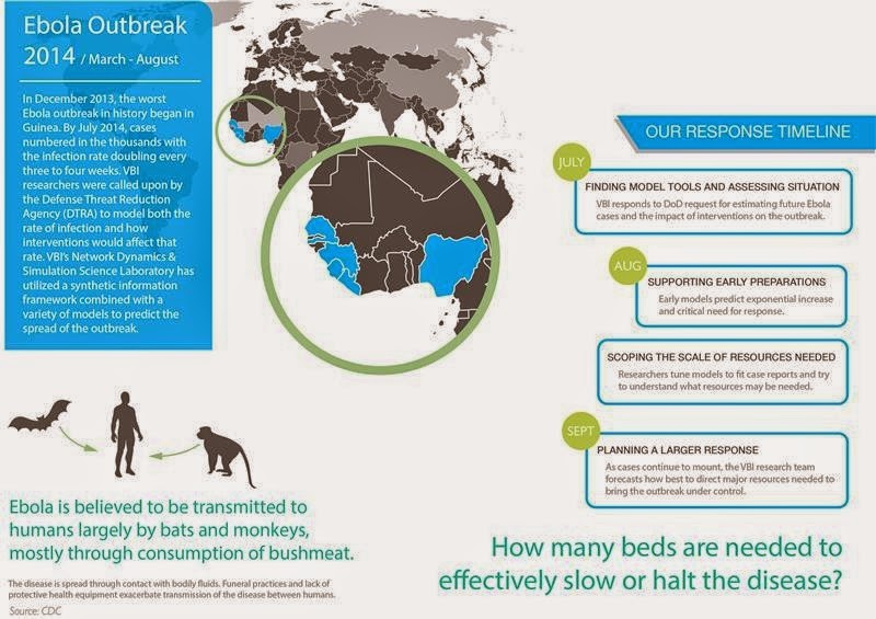
The Network Dynamics and Simulation Science Laboratory at the Virginia Bioinformatics Institute modeled the rate of infections and how interventions would affect the rate.
Credit: Source: CDC / Image courtesy of Virginia Tech
The Ebola epidemic could claim hundreds of thousands of lives and infect more than 1.4 million people by the end of January, according to a statistical forecast released this week by the U.S. Centers for Disease Control and Prevention.

The CDC forecast supports the drastically higher projections released earlier by a group of scientists, including epidemiologists with the Virginia Bioinformatics Institute, who modeled the Ebola spread as part of a National Institutes of Health-sponsored project called Midas, short for Models of Infectious Disease Agent Study.

The effort is also supported by the federal Defense Threat Reduction Agency.
Before the scientists released results, the outbreak in West Africa was expected to be under control in nine months with only about 20,000 total cases. But modeling showed 20,000 people could be infected in just a single month.

The predictions could change dramatically if public health efforts become effective, but based on the virus's current uncontrolled spread, numbers of people infected could skyrocket.

"If the disease keeps spreading as it has been we estimate there could be hundreds of thousands of cases by the end of the year in Liberia alone," said Bryan Lewis, a computational epidemiologist with the Network Dynamics and Simulation Science Laboratory at the Virginia Bioinformatics Institute.

Lewis and his fellow researchers use a combination of models to predict outcomes of the epidemic.

The agent-based models are adaptive, evolving as more information is fed into them to provide an accurate forecast.

Pharmaceutical intervention, which is still on the horizon, is proving less effective in the models than supportive care and personal protection equipment for health care workers.

"The work with Ebola is not an isolated event," said Christopher Barrett, the executive director of the institute. "This research is part of a decades-long effort largely funded by the Defense Threat Reduction Agency to build a global synthetic population that will allow us to ask questions about our world and ourselves that we have never been able to ask before, and to use those answers to prevent or quickly intervene during a crisis."

Barrett and other institute leaders updated U.S. Sen. Tim Kaine and Virginia Tech President Timothy Sands about the Network Dynamics and Simulation Science Lab's role in analyzing the Ebola outbreak at the Virginia Tech Research Center in Arlington on Tuesday morning. That afternoon in Blacksburg they briefed staff members from U.S. Sen. Mark Warner's office.

A university-level Research Institute of Virginia Tech, the Virginia Bioinformatics Institute was established in 2000 with an emphasis on informatics of complex interacting systems scaling the microbiome to the entire globe. It helps solve challenges posed to human health, security, and sustainability. Headquartered at the Blacksburg campus, the institute occupies 154,600 square feet in research facilities, including state-of-the-art core laboratory and high-performance computing facilities, as well as research offices in the Virginia Tech Research Center in Arlington, Virginia.

Source: Virginia Tech

Up to 80 million bacteria sealed with a kiss

Written By Unknown on Tuesday, January 6, 2015 | 1:57 AM

Couple about to kiss (stock image). As many as 80 million bacteria are transferred during a 10 second kiss, according to research published in the open access journal Microbiome.
As many as 80 million bacteria are transferred during a 10 second kiss, according to research published in the open access journal Microbiome. The study also found that partners who kiss each other at least nine times a day share similar communities of oral bacteria.

The ecosystem of more than 100 trillion microorganisms that live in our bodies -- the microbiome -- is essential for the digestion of food, synthesizing nutrients, and preventing disease. It is shaped by genetics, diet, and age, but also the individuals with whom we interact. With the mouth playing host to more than 700 varieties of bacteria, the oral microbiota also appear to be influenced by those closest to us.

Researchers from Micropia and TNO in the Netherlands studied 21 couples, asking them to fill out questionnaires on their kissing behaviour including their average intimate kiss frequency. They then took swab samples to investigate the composition of their oral microbiota on the tongue and in their saliva.

The results showed that when couples intimately kiss at relatively high frequencies their salivary microbiota become similar. On average it was found that at least nine intimate kisses per day led to couples having significantly shared salivary microbiota.

Lead author Remco Kort, from TNO's Microbiology and Systems Biology department and adviser to the Micropia museum of microbes, said: "Intimate kissing involving full tongue contact and saliva exchange appears to be a courtship behavior unique to humans and is common in over 90% of known cultures. Interestingly, the current explanations for the function of intimate kissing in humans include an important role for the microbiota present in the oral cavity, although to our knowledge, the exact effects of intimate kissing on the oral microbiota have never been studied. We wanted to find out the extent to which partners share their oral microbiota, and it turns out, the more a couple kiss, the more similar they are."

In a controlled kissing experiment to quantify the transfer of bacteria, a member of each of the couples had a probiotic drink containing specific varieties of bacteria including Lactobacillus and Bifidobacteria. After an intimate kiss, the researchers found that the quantity of probiotic bacteria in the receiver's saliva rose threefold, and calculated that in total 80 million bacteria would have been transferred during a 10 second kiss.

The study also suggests an important role for other mechanisms that select oral microbiota, resulting from a shared lifestyle, dietary and personal care habits, and this is especially the case for microbiota on the tongue. The researchers found that while tongue microbiota were more similar among partners than unrelated individuals, their similarity did not change with more frequent kissing, in contrast to the findings on the saliva microbiota.

Commenting on the kissing questionnaire results, the researchers say that an interesting but separate finding was that 74% of the men reported higher intimate kiss frequencies than the women of the same couple. This resulted in a reported average of ten kisses per day from the males, twice that of the female reported average of five per day.

To calculate the number of bacteria transferred in a kiss, the authors relied on average transfer values and a number of assumptions related to bacterial transfer, the kiss contact surface, and the value for average saliva volume.

How llamas' unusual antibodies might help in the fight against HIV/AIDS

Written By Unknown on Sunday, December 28, 2014 | 7:15 PM

Llamas contribute to the fight against AIDS. Credit: Nika Stropakke, CC-BY
Most vaccines work by inducing an immune response characterized by neutralizing antibodies against the respective pathogen. An effective HIV vaccine has remained elusive so far, but researchers have continued to make progress, often employing innovative methods. A study published on December 18th in PLOS Pathogens reports that a combination of antibodies from llamas can neutralize (destroy) a wide range of circulating HIV viruses.

After initial disappointment that HIV vaccine candidates were unable to elicit neutralizing antibodies, researchers found that some HIV-infected individuals did produce such antibodies. The current challenge is therefore to find safe and effective vaccine formulations (as opposed to HIV infection) that trigger the development of neutralizing antibodies that can recognize and prevent infection with all or most circulating HIV subtypes.

Many known neutralizing antibodies are directed against a specific part of the virus that binds to the CD4 receptor on the human target cells, and structural biology studies indicated that the site is a narrow groove. Antibodies in most mammals are relatively large proteins made up of two copies of two different individual parts (or chains), and bulkiness might be one reason why neutralizing antibodies are rare. Llamas are a notable exception: besides the common four-chain antibodies they also produce smaller ones made up of only two of the four chains. Robin Weiss, an HIV expert, and Theo Verrips, a llama antibody expert, therefore started working with this unconventional research animal.

Laura McCoy (working with Weiss at University College London, UK) led an international group of researchers to test immunization protocols and the resulting immune response in llamas. Having previously identified one particular HIV neutralizing llama antibody, for this study the researchers immunized two additional llamas and identified a total of three new neutralizing antibodies. The four HIV neutralizing llama antibodies target different parts of the CD4-binding site of the virus, and the researchers could show that when used in combination, rather than interfering with each other, they are more potent and can neutralize all of the 60 different HIV strains tested.

To understand how the llama immunization--which included two sets of four sequential vaccine injections per animal--worked, the researchers sequenced many copies of antibody-coding genes from blood cells collected after the first set of immunizations and after a further four rounds of vaccination. They also looked at the "naïve" antibody repertoire from seven llamas that had not been vaccinated. The results suggest that the neutralizing antibodies were not part of the pre-immunization repertoire, nor were they detectable after the first vaccination round. Rather, they were generated as immune cells repeatedly encountered the vaccine and responded by maturing specific antibodies that can recognize it.

While it is encouraging that broadly neutralizing antibodies were found in all of the immunized llamas, they are present only at low concentrations in the blood, and so fail to meet the goal for a protective HIV vaccine. Nonetheless, the researchers conclude that the llama model has allowed them to examine the generation of four broadly neutralizing antibodies induced by vaccination, which has not been possible in any other species.

Source: PLOS.

Infectious prion protein discovered in urine of patients with variant Creutzfeldt-Jakob disease

Claudio Soto, Ph.D., in one of his labs at The University of Texas Health Science Center at Houston (UTHealth). Credit: Image courtesy of University of Texas Health Science Center at Houston
The misfolded and infectious prion protein that is a marker for variant Creutzfeldt-Jakob disease – linked to the consumption of infected cattle meat – has been detected in the urine of patients with the disease by researchers at The University of Texas Health Science Center at Houston (UTHealth) Medical School.

The results of the international study, led by Claudio Soto, Ph.D., professor of neurology at the UTHealth Medical School, will be published in the Aug. 7 issue of the New England Journal of Medicine.

Variant Creutzfeldt-Jakob disease in humans and bovine spongiform encephalopathy in animals – also known as Mad Cow disease – are fatal neurodegenerative disorders. There are currently no noninvasive tools available to diagnose the disease and there are no treatments.

Sporadic Creutzfeldt-Jakob disease occurs worldwide at a rate of around 1 new case per million people per year. The variant form is a new disease occurring in people who either ate the beef of cows with bovine spongiform encephalopathy or, in the case of three patients in the United Kingdom, received blood transfusions from asymptomatic infected donors.

The international team of researchers analyzed urine samples from 68 patients with sporadic Creutzfeldt-Jakob disease, 14 patients with variant Creutzfeldt-Jakob disease, four patients with genetic prion diseases, 50 patients with other neurodegenerative diseases, 50 patients with nondegenerative neurologic diseases and 52 healthy persons.

Soto’s laboratory used a protein misfolding cyclic amplification assay, invented in the lab, which mimics the prion replication process in vitro that occurs in prion disease. The misfolded prion proteins were detected in the urine of 13 of 14 patients with variant Creutzfeldt-Jakob disease. The single patient whose urine was negative had been receiving an experimental treatment of pentosan polysulfate directly into the brain. No misfolded prion proteins were detected in the urine of any the other study subjects, including the patients who had sporadic Creutzfeldt-Jakob disease.

“What could be less invasive than detecting this disease in urine? The fact that we were able to detect just the variant Creutzfeldt-Jakob disease form in the urine is very important. This could lead to the development of commercial technology for diagnosis as well as to determine the safety of donated blood and urinary products,” said Soto, who is the director of The George and Cynthia Mitchell Center for Research in Alzheimer’s disease and Related Brain Disorders, and founder of Amprion Inc, a biotech company developing the cyclic amplification technology for commercial application.

According to the World Health Organization (WHO), variant Creutzfeldt-Jakob disease affects younger patients, who have a median age of 28 at death, compared to sporadic Creutzfeldt-Jakob disease with a median age of 68. Most patients, after diagnosis of either form, live less than a year before death.

As of June 2, 2014, 177 of 229 people in the world with Creutzfeldt-Jakob disease were from the United Kingdom. A 2013 study published in the British Medical Journal has estimated that approximately 30,000 people in the United Kingdom might be carriers of the variant form of the disease.

“This study reports, for the first time, the detection of the abnormal prion protein in the urine from patients with variant Creutzfeldt-Jakob disease using the protein misfolding amplification technique pioneered by Dr. Claudio Soto,” said co-author James W. Ironside, FMedSci, FRSE, professor of clinical neuropathology at the National CJD Research and Surveillance Unit at the University of Edinburgh. “This has great potential to allow the development of a highly sensitive and specific non-invasive test that can be used for the diagnosis of variant Creutzfeldt-Jakob disease, and potentially as a screening tool for variant Creutzfeldt-Jakob disease infection in asymptomatic individuals, which is a topic of current interest in the United Kingdom.”

Source: University of Texas Health Science Center at Houston

Bacteria could be rich source for making terpenes

Written By Unknown on Friday, December 26, 2014 | 4:18 PM

Odoriferous terpene metabolites: A phylogenetic tree of terpene synthases shows the synthases (bold face or underlined) found by researchers in Japan and at Brown University using bacterial sequences. Credit: Image courtesy of Brown University
If you've ever enjoyed the scent of a pine forest or sniffed a freshly cut basil leaf, then you're familiar with terpenes. The compounds are responsible for the essential oils of plants and the resins of trees. Since the discovery of terpenes more than 150 years ago, scientists have isolated some 50,000 different terpene compounds derived from plants and fungi. Bacteria and other microorganisms are known to make terpenes too, but they've received much less study.

New research at Brown University, published in the Proceedings of the National Academy of Sciences, shows that the genetic capacity of bacteria to make terpenes is widespread. 

Using a specialized technique to sift through genomic databases for a variety of bacteria, the researchers found 262 gene sequences that likely code for terpene synthases -- enzymes that catalyze the production terpenes. The researchers then used several of those enzymes to isolate 13 previously unidentified bacterial terpenes.

The findings suggest that bacteria "represent a fertile source for discovery of new natural products," the researchers write.

David Cane, a professor of chemistry at Brown and one of the authors on the new paper, began working about 15 years ago to understand how bacteria make terpenes.

"At that time, the first genomic sequences of certain classes of bacteria were just beginning to come out," he said. "We had this idea that maybe you could find the enzymes responsible for making terpenes by looking at the sequences of the genes that were being discovered."

To do that, Cane searched through the genome data gathered for a group of bacteria called Streptomyces, looking for sequences similar those known to produce terpene synthases in plants and fungi. Eventually, he found that Streptomyces did indeed have genes encoding terpene synthases and that those enzymes could be used to make terpenes.

The verified bacterial sequences found by Cane and others enabled researchers to refine subsequent searches for additional terpene synthase genes. "Instead of using plant sequences or fungal sequences as your search query, we can now use bacterial sequences, which should yield a greater degree of similarity," he said. "So now we're fishing in the right waters with the right kind of bait, and you can find more matches."

This latest paper made use of the third generation of iterative searches and a powerful search technique developed by Haruo Ikeda of Kitasato University in Japan. Previous work had identified 140 probable sequences for terpene synthases. This latest work expanded that to 262.

The next step was to verify that these sequences did indeed code for enzymes capable of making terpenes. Testing all 262 wasn't practical, so the team chose a few they thought might give them the best chance of finding terpene compounds that hadn't previously been identified. They looked for sequences that didn't seem to fit clearly into previously known categories of terpenes.

After they had selected a few, the team made use of a genetically engineered Streptomyces bacterium as a bio-refinery to generate the terpene products.

"What Professor Ikeda did, in collaboration with us, is develop a variant of a very well-studied Streptomyces system," Cane said. "He eliminated the genes that were responsible for making most of its native products, but he left behind all of the capacity to provide the starting materials and handle the accumulation of products."

By taking some of the gene sequences they found and splicing them into their test organism, the researchers could let the organisms generate the product using the instructions from the newly introduced gene. Using this method, they were able to make 13 previously unknown terpenes, their structures verified by mass spectrometry and nuclear magnetic resonance spectroscopy.

"It's a big step forward in the area in that it provides a paradigm for how one could go about discovering many new substances," Cane said. "It's a good example of how one can use sequence analysis to identify genes of interest and then apply molecular genetic and microbiological techniques to produce the chemical substances of interest."

The work also suggests that there may be many new terpene products as yet undiscovered hiding in the genomes of bacteria.

Source: Brown University

Role of microbes, effect on infectious disease dynamics

Written By Unknown on Wednesday, December 24, 2014 | 7:24 PM

In the field, UCSB graduate student Andrea Jani uses a sterile swab to sample mucus on the skin of a frog. Frogs are released unharmed after sampling. Credit: Abby Mayer
The adult human body is made up of about 37 trillion cells. Microbes, mainly bacteria, outnumber body cells by 10 to 1. Increasingly, scientists recognize that this huge community of microbes, called the microbiome, affects the health, development and evolution of all multicellular organisms, including humans.

Studies show symbiotic microbes can help prevent infection by disease-causing pathogens. But sometimes the interaction goes the other way, with a pathogen or disease disrupting the normal community of symbiotic bacteria. In a new study, a team of scientists from UC Santa Barbara demonstrates that a fungal pathogen of amphibians does just that. The findings appear today in the Early Edition of the Proceedings of the National Academy of Science.

Landmark experiments with model organisms such as mice have shown that infectious pathogens can disrupt the "normal" microbiome, but the extent to which this process shapes symbiotic microbial communities during disease outbreaks in nature is largely unknown. This new work, conducted by Andrea Jani, a UCSB graduate student in Cherie Briggs' lab in the Department of Ecology, Evolution and Marine Biology (EEMB), addresses a fundamental gap in disease ecology and microbiome research.

Co-authors Jani and Briggs -- also affiliated with UCSB's Biomolecular Science and Engineering program -- found that the chytrid fungus Batrachochytrium dendrobatidis (Bd) appears to drive dramatic changes in symbiotic bacterial communities during natural disease episodes in four populations of the endangered Sierra Nevada yellow-legged frog (Rana sierrae). Chytridiomycosis, an emerging infectious disease of amphibian skin caused by the Bd pathogen, is a leading cause of amphibian biodiversity loss worldwide.

"In the California Sierra Nevada, this disease has led to the rapid extirpation of frogs from hundreds of high-elevation lakes; however, in other lakes, infected frogs of the same species are surviving and persisting with the fungus," explained Briggs, who is the Duncan and Suzanne Mellichamp Chair in Systems Biology. "Given that amphibian skin is the organ infected by Bd, there has been a lot of interest in how antifungal properties of some skin-associated bacteria may protect frogs against this fungal pathogen. In this study we focused on the flip side of this interaction -- that is, how infection with Bd can disrupt the skin microbial community."

"We used next-generation DNA sequencing to document significant shifts in skin-associated bacterial communities of the Sierra Nevada yellow-legged frog during natural Bd outbreaks," Jani explained. "We paired these field surveys with a laboratory infection experiment, demonstrating a causal relationship in which Bd alters the Rana sierrae microbiome."

The researchers found a remarkable consistency in the response of the microbiome to Bd infection among field populations and between the field and laboratory. Several key taxa -- a group of one or more populations of an organism or organisms -- consistently responded in the same direction to Bd infection, suggesting some predictability in the effect of Bd on the microbiome.

"What we found was that the severity of infection with Bd is strongly correlated with the composition of bacterial communities on the skin of frogs," Jani continued. "What was surprising was that across the different frog populations there was pretty striking consistency in this correlation with Bd. One of the frog populations crashed due to Bd infection; the other three populations seemed to tolerate Bd infections. So there are different disease dynamics going on, yet they have a similar relationship between the microbiome and Bd."

Still, the underlying mechanism for Bd-induced changes in the microbiome is not clear. The researchers hypothesize that the pathogen might compete directly with certain bacteria for space or resources or release compounds that negatively or positively affect certain bacterial species. Alternatively, they say, some pathogens could control immune responses of the host to favor their own growth and disrupt the normal symbiotic bacterial community.

Jani noted that some promise exists for probiotic treatments as a tool to fight the decline of frogs due to Bd, but she was careful to qualify that statement by saying that there is still a lot that scientists do not understand about either the environmental impact that might have or what the interactions are between the natural bacteria that exist on frogs and the pathogen. "We find that some taxa previously identified as having anti-Bd properties are driven to low abundances by Bd infection, which may limit their effectiveness as probiotic agents," she said.

"This study shows the importance of knowing how the many benign microbes living on and in our bodies interact with those that cause disease," said Sam Scheiner, National Science Foundation (NSF) director for the joint NSF/National Institutes of Health/United States Department of Agriculture Ecology and Evolution of Infectious Disease Program, which funded the research. "The results are important for developing responses to a disease causing amphibians to go extinct worldwide and also have implications for future studies of human health.

Source:  University of California - Santa Barbara
 
Support : Creating Website | Johny Template | Mas Template
Copyright © 2011. The planet wall - All Rights Reserved
Template Created by Easy Blogging Published by Mas Template
Proudly powered by Blogger