Latest Post
Showing posts with label PANDEMIC. Show all posts

Computational model: Ebola could infect more than 1.4 million people by end of January 2015

Written By Unknown on Wednesday, January 7, 2015 | 7:21 AM

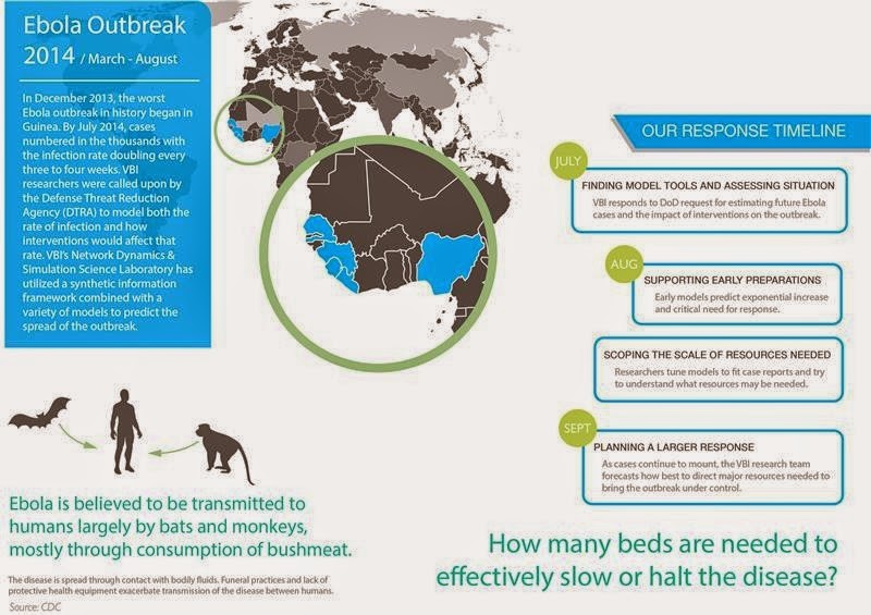
The Network Dynamics and Simulation Science Laboratory at the Virginia Bioinformatics Institute modeled the rate of infections and how interventions would affect the rate.
Credit: Source: CDC / Image courtesy of Virginia Tech
The Ebola epidemic could claim hundreds of thousands of lives and infect more than 1.4 million people by the end of January, according to a statistical forecast released this week by the U.S. Centers for Disease Control and Prevention.

The CDC forecast supports the drastically higher projections released earlier by a group of scientists, including epidemiologists with the Virginia Bioinformatics Institute, who modeled the Ebola spread as part of a National Institutes of Health-sponsored project called Midas, short for Models of Infectious Disease Agent Study.

The effort is also supported by the federal Defense Threat Reduction Agency.
Before the scientists released results, the outbreak in West Africa was expected to be under control in nine months with only about 20,000 total cases. But modeling showed 20,000 people could be infected in just a single month.

The predictions could change dramatically if public health efforts become effective, but based on the virus's current uncontrolled spread, numbers of people infected could skyrocket.

"If the disease keeps spreading as it has been we estimate there could be hundreds of thousands of cases by the end of the year in Liberia alone," said Bryan Lewis, a computational epidemiologist with the Network Dynamics and Simulation Science Laboratory at the Virginia Bioinformatics Institute.

Lewis and his fellow researchers use a combination of models to predict outcomes of the epidemic.

The agent-based models are adaptive, evolving as more information is fed into them to provide an accurate forecast.

Pharmaceutical intervention, which is still on the horizon, is proving less effective in the models than supportive care and personal protection equipment for health care workers.

"The work with Ebola is not an isolated event," said Christopher Barrett, the executive director of the institute. "This research is part of a decades-long effort largely funded by the Defense Threat Reduction Agency to build a global synthetic population that will allow us to ask questions about our world and ourselves that we have never been able to ask before, and to use those answers to prevent or quickly intervene during a crisis."

Barrett and other institute leaders updated U.S. Sen. Tim Kaine and Virginia Tech President Timothy Sands about the Network Dynamics and Simulation Science Lab's role in analyzing the Ebola outbreak at the Virginia Tech Research Center in Arlington on Tuesday morning. That afternoon in Blacksburg they briefed staff members from U.S. Sen. Mark Warner's office.

A university-level Research Institute of Virginia Tech, the Virginia Bioinformatics Institute was established in 2000 with an emphasis on informatics of complex interacting systems scaling the microbiome to the entire globe. It helps solve challenges posed to human health, security, and sustainability. Headquartered at the Blacksburg campus, the institute occupies 154,600 square feet in research facilities, including state-of-the-art core laboratory and high-performance computing facilities, as well as research offices in the Virginia Tech Research Center in Arlington, Virginia.

Source: Virginia Tech

Trial confirms Ebola vaccine candidate safe, equally immunogenic in Africa

Written By Unknown on Sunday, December 28, 2014 | 7:35 PM

"This is the first study to show comparable safety and immune response of an experimental Ebola vaccine in an African population," says lead author Dr Julie Ledgerwood. "This is particularly encouraging because those at greatest risk of Ebola live primarily in Africa, and diminished vaccine protection in African populations has been seen for other diseases." Credit: © nito / Fotolia
Two experimental DNA vaccines to prevent Ebola virus and the closely related Marburg virus are safe, and generated a similar immune response in healthy Ugandan adults as reported in healthy US adults earlier this year. The findings, from the first trial of filovirus vaccines in Africa, are published in The Lancet.

"This is the first study to show comparable safety and immune response of an experimental Ebola vaccine in an African population," says lead author Dr Julie Ledgerwood from the National Institutes of Allergy and Infectious Diseases (NIAID) at the National Institutes of Health, USA. "This is particularly encouraging because those at greatest risk of Ebola live primarily in Africa, and diminished vaccine protection in African populations has been seen for other diseases."

Scientists from the NIAID developed the DNA vaccines that code for Ebola virus proteins from the Zaire and Sudan strains and the Marburg virus protein. The vaccines contain the construction plans for the proteins on the outer surface of the virus. Immune responses against these proteins have shown to be highly protective in non-human primate models.

In this phase 1 trial, the Makerere University Walter Reed Program enrolled 108 healthy adults aged between 18 and 50 from Kampala, Uganda between November, 2009 and April, 2010. Each volunteer was randomly assigned to receive an intramuscular injection of either the Ebola vaccine (30 volunteers), Marburg vaccine (30), both vaccines (30), or placebo (18) at the start of the study, and again 4 weeks and 8 weeks later.

The vaccines given separately and together were safe and stimulated an immune response in the form of neutralising antibodies and T-cells against the virus proteins. Four weeks after the third injection, just over half of the volunteers (57%; 17 of 30) had an antibody response to the Ebola Zaire protein as did 14 of 30 participants who received both the Ebola and Marburg vaccines. 

However, the antibodies were not long-lasting and returned to undetectable levels within 11 months of vaccination.

Both DNA vaccines were well tolerated in Ugandan adults with similar numbers of local and systemic reactions reported in all groups. Only one serious adverse event (neutropenia; low white blood cell count) was reported in a Marburg vaccine only recipient, but was not thought to be vaccine related.

According to Dr Ledgerwood, "These findings have already formed the basis of a more potent vaccine, delivered using a harmless chimpanzee cold virus, which is undergoing trials in the USA, UK, Mali, and Uganda in response to the ongoing Ebola virus outbreak."

Writing in a linked Comment, Dr Saranya Sridhar from the Jenner Institute at the University of Oxford in the UK says, "[This] study deserves to be the focal point around which the broader question of vaccine development, particularly for Africa, must be addressed. With the uncharitable benefit of hindsight in view of the evolving 2014 Ebola outbreak, we must ask ourselves whether a filovirus vaccine should have been in more advanced clinical development. The international response to the present Ebola outbreak is an exemplar of the speed and purpose with which clinical vaccine development can progress and has set the benchmark against which future vaccine development must be judged. This study is the first step on the aspirational road towards the deployment of filovirus vaccines in Africa and must serve to shake the metaphorical cobwebs that can stall our advance towards this destination."

Source: The Lancet

Scrapie could breach the species barrier

Written By Unknown on Thursday, December 25, 2014 | 3:05 AM

Scrapie is a neurodegenerative disease that has been known for centuries and which affects sheep and goats. Credit: INRA/Florent Giffard
INRA scientists have shown for the first time that the pathogens responsible for scrapie in small ruminants (prions) have the potential to convert the human prion protein from a healthy state to a pathological state. In mice models reproducing the human species barrier, this prion induces a disease similar to Creutzfeldt-Jakob disease. These primary results published in Nature Communications on 16 December 2014, stress the necessity to reassess the transmission of this disease to humans.

Scrapie is a neurodegenerative disease that has been known for centuries and which affects sheep and goats. Similar to Bovine Spongiform Encephalopathy (BSE) or mad cow disease, scrapie is caused by a transmissible pathogen protein called prion.

However, and contrary to BSE[1], epidemiological studies have never been able to establish a link between this disease and the occurrence of prion diseases in humans. "Risks of transmitting scrapie to humans (zoonose) were hitherto considered negligible because of the species barrier that naturally prevents prion propagation between species," said Olivier 
Andreoletti, INRA scientist who led the present study.

Researchers at INRA studied the permeability of the human transmission barrier to pathogens responsible for scrapie, using animal models specifically developed for this purpose. This approach previously allowed the confirmation of the zoonotic nature of prions responsible for BSE in cows and of the variant of Creutzfeldt-Jakob disease in humans (vCJD).

Unexpectedly, in these rodent models, certain pathogens responsible for scrapie were able to cross the transmission barrier. Moreover, the pathogens that propagated through this barrier were undistinguishable from the prions causing the sporadic form of Creutzfeldt-Jakob disease (sCJD). This data suggest a potential link between the occurrence of certain sCJD and these animal prions.

"Since CJD is scarce, about 1 case per million and per year, and incubation periods are usually long -several decades- it is extremely difficult for epidemiological studies to try and make this link," explains Olivier Andreoletti.

In their conclusions, the authors stress the fact that CJD cases are rare though scrapie has been circulating for centuries in small ruminants for which we eat the meat. Even if in future studies scrapie is finally confirmed to have a zoonotic potential, the authors consider that this disease does not constitute a new major risk for public health.

Source: INRA-France

Genetic factors behind surviving or dying from Ebola shown in mouse study

In an emerging disease research lab at the University of Washington, Chris Williams, a research scientist who specializes in microbiology laboratory robotics, programs a piece of equipment that can be programmed to performs many lab tasks. Credit: Brian Donohue
A newly developed mouse model suggests that genetic factors are behind the mild-to-deadly range of reactions to the Ebola virus.

People exposed to Ebola vary in how the virus affects them. Some completely resist the disease, others suffer moderate to severe illness and recover, while those who are most susceptible succumb to bleeding, organ failure and shock.
In earlier studies of populations of people who have contracted Ebola, these differences are not related to any specific changes in the Ebola virus itself that made it more or less dangerous; instead, the body's attempts to fight infection seems to determine disease severity.

In the Oct. 30 edition of Science, scientists describe strains of laboratory mice bred to test the role of an individual's genetic makeup in the course of Ebola disease. Systems biologists and virologists Angela Rasmussen and Michael Katze from the Katze Laboratory at the University of Washington Department of Microbiology led the study in collaboration with the National Institutes of Health's Rocky Mountain Laboratories in Montana and University of North Carolina at Chapel Hill.

Research on Ebola prevention and treatment has been hindered by the lack of a mouse model that replicates the main characteristics of human Ebola hemorrhagic fever. The researchers had originally obtained this genetically diverse group of inbred laboratory mice to study locations on mouse genomes associated with influenza severity.

The research was conducted in a highly secure, state-of-the-art biocontainment safety level 4 laboratory in Hamilton, Mont. The scientists examined mice that they infected with a mouse form of the same species of Ebola virus causing the 2014 West Africa outbreak. The study was done in full compliance with federal, state, and local safety and biosecurity regulations. This type of virus has been used several times before in research studies. Nothing was done to change the virus.

Interestingly, conventional laboratory mice previously infected with this virus died, but did not develop symptoms of Ebola hemorrhagic fever.

In the present study, all the mice lost weight in the first few days after infection. Nineteen percent of the mice were unfazed. They not only survived, but also fully regained their lost weight within two weeks. They had no gross pathological evidence of disease. Their livers looked normal.

Eleven percent were partially resistant and less than half of these died. Seventy percent of the mice had a greater than 50 percent mortality. Nineteen percent of this last group had liver inflammation without classic symptoms of Ebola, and thirty-four percent had blood that took too long to clot, a hallmark of fatal Ebola hemorrhagic fever in humans. Those mice also had internal bleeding, swollen spleens and changes in liver color and texture.
The scientists correlated disease outcomes and variations in mortality rates to specific genetic lines of mice.

"The frequency of different manifestations of the disease across the lines of these mice screened so far are similar in variety and proportion to the spectrum of clinical disease observed in the 2014 West African outbreak," Rasmussen said.

While acknowledging that recent Ebola survivors may have had immunity to this or a related virus that saved them during this epidemic, Katze said, "Our data suggest that genetic factors play a significant role in disease outcome."

In general, when virus infection frenzied the genes involved in promoting blood vessel inflammation and cell death, serious or fatal disease followed. On the other hand, survivors experienced more activity in genes that order blood vessel repair and the production of infection-fighting white blood cells.

The scientists note that certain specialized types of cells in the liver could also have limited virus reproduction and put a damper on systemic inflammation and blood clotting problems in resistant mice. Susceptible mice had widespread liver infection, which may explain why they had more virus in their bodies and poorly regulated blood coagulation. The researchers also noticed that spleens in the resistant and susceptible mice took alternate routes to try to ward off infection.

"We hope that medical researchers will be able to rapidly apply these findings to candidate therapeutics and vaccines," Katze said. They believe this mouse model can be promptly implemented to find genetic markers, conduct meticulous studies on how symptoms originate and take hold, and evaluate drugs and that have broad spectrum anti-viral activities against all Zaire ebolaviruses, including the one responsible for the current West African epidemic.

For tiger populations, a new threat

Written By Unknown on Wednesday, December 24, 2014 | 5:23 AM

This is an Amur tiger photographed by camera trap. Credit: WCS Russia Program
Along with the pressures of habitat loss, poaching and depletion of prey species, a new threat to tiger populations in the wild has surfaced in the form of disease, specifically, canine distemper virus (CDV). According to a new study from the Wildlife Conservation Society (WCS) and its partners, CDV has the potential to be a significant driver in pushing the animals toward extinction.

While CDV has recently been shown to lead to the deaths of individual tigers, its long-term impacts on tiger populations had never before been studied.

The authors evaluated these impacts on the Amur tiger population in Russia's Sikhote-Alin Biosphere Zapovednik (SABZ), where tiger numbers declined from 38 individuals to 9 in the years 2007 to 2012. In 2009 and 2010, six adult tigers died or disappeared from the reserve, and CDV was confirmed in two dead tigers -- leading scientists to believe that CDV likely played a role in the overall decline of the population. Joint investigations of CDV have been an ongoing focus of WCS and Russian scientists at Sikhote-Alin Zapovednik and veterinarians at the regional Primorye Agricultural College since its first appearance in tigers in 2003.

A key finding of this study: Modeling shows that smaller populations of tigers were found to be more vulnerable to extinction by CDV. Populations consisting of 25 individuals were 1.65 times more likely to decline in the next 50 years when CDV was present. The results are profoundly disturbing for global wild tigers given that in most sites where wild tigers persist they are limited to populations of less than 25 adult breeding individuals.

The scientists used computer modeling to simulate the effects of CDV infection on isolated tiger populations of various sizes and through a series of transmission scenarios. These included tiger-to-tiger transmission and transmission through predation on CDV-infected domestic dogs and/or infected wild carnivores (such as foxes, raccoon dogs and badgers). High and low-risk scenarios for the model were created based on variation in the prevalence of CDV and the tigers' contact with sources of exposure.

Results showed that CDV infection increased the 50-year extinction probability of tigers in SABZ as much as 55.8 percent compared to CDV-free populations of equivalent size.

"Although we knew that individual tigers had died from CDV in the wild, we wanted to understand the risk the virus presents to whole populations," said WCS veterinarian Martin Gilbert. "Tigers are elusive, however, and studying the long-term impact of risk factors is very challenging. Our model, based on tiger ecology data collected over 20 years in SABZ, explored the different ways that tigers might be exposed to the virus and how these impact the extinction risk to tiger populations over the long term."

WCS Russia Program Director Dale Miquelle said, "Tigers face an array of threats throughout their range, from poaching to competition with humans for space and for food. 

Consequently, many tiger populations have become smaller and more fragmented, making them much more susceptible to diseases such as CDV. While we must continue to focus on the primary threats of poaching and habitat destruction, we now must also be prepared to deal with the appearance of such diseases in the future."

Priorities for future research, according to the authors, include identifying the domestic and wild carnivore species that contribute to the CDV reservoir, and those that are the most likely sources of infection for tigers. Tigers are too rare to sustain the virus in the long term, so CDV must rely on more abundant carnivore species to persist in the environment. 

Understanding the structure of the CDV reservoir will be a critical first step in identifying measures that might prevent or control future outbreaks. In addition, since we now know that small tiger populations are at greater risk to diseases such as CDV than larger populations, conservation strategies focusing on connectedness between populations become all the more important. "Estimating the potential impact of canine distemper virus on the Amur tiger population (Panthera tigris altaica) in Russia," appears in the current online edition of PloSONE. Authors include: Martin Gilbert of WCS and Boyd Orr Centre for Population and Ecosystem Health at the University of Glasgow; Dale G. Miquelle of WCS; John M. Goodrich of Panthera; Richard Reeve, Sarah Cleaveland and Louise Matthews of Boyd Orr Centre for Population and Ecosystem Health at the University of Glasgow; and Damien Joly of WCS and Metabiota.

This study was made possible through generous support from Morris Animal Foundation, Zoo Boise Conservation Fund, AZA Tiger Species Survival Plan Tiger Conservation Campaign, and the Biotechnology and Biological Sciences Research Council.

"Morris Animal Foundation is thankful to Dr. Miquelle and his team for helping protect the Amur Tigers," said Diane Brown, DVM, PhD, DACVP and Chief Scientific Officer for Morris Animal Foundation. "Our Foundation values their hard work and dedication to this study and we look forward to many more partnerships with the Wildlife Conservation Society."

Source: Wildlife Conservation Society

Using power of computers to harness human genome may provide clues into Ebola virus

Written By Unknown on Tuesday, December 23, 2014 | 5:39 PM

Ramaswamy Narayanan, Ph.D., professor in the Charles E. Schmidt College of Science at Florida Atlantic University.
Ramaswamy Narayanan, Ph.D., professor in the Charles E. Schmidt College of Science at Florida Atlantic University, is working to blend the power of computers with biology to use the human genome to remove much of the guesswork involved in discovering cures for diseases.

In an article titled "Ebola-Associated Genes in the Human Genome: Implications for Novel Targets," published in the current MedCrave Online Journal of Proteomics and Bioinformatics, Narayanan describes how key genes that are present in our cells could be used to develop drugs for this disease.

"Bioinformatics is a powerful tool to help us understand biological data," said Narayanan whose research has focused in this field for more than a decade. "We are mining the human genome for Ebola virus association to develop an understanding of the human proteins involved in this disease for subsequent research and development, and to potentially create a pipeline of targets that we can test and evaluate."

Ebola virus disease is a major healthcare challenge facing the globe today and if left unchecked could become a pandemic. A limited knowledgebase exists about the Ebola virus and companies are hastening to develop vaccines and other forms to treat and cure the virus. There are no FDA-approved drugs, and developing vaccines or antibodies and testing them in clinical trials is an arduous process that takes considerable time. Currently, patients infected with Ebola are only able to receive supportive care such as fluid replacement, nutritional support, pain control, and blood pressure maintenance. In some cases, patients may be fortunate enough to be treated with experimental drugs.

Narayanan's work has helped to identify numerous FDA-approved drugs already used for many other diseases including anti-inflammatory drugs, anticoagulants, cancer, HIV, statins and hormones, which could potentially be used to add to the current supportive care for patients with the Ebola virus.

"With the high mortality rate of this disease, the world urgently needs new ways to treat patients," said Narayanan. "The ability to use drugs that are already approved by the FDA could provide clinicians with more options to treat Ebola patients, rather than just relying on supportive measures like fluid replacement or antibiotics."

According to the World Health Organization (WHO), Ebola virus disease (EVD) is a severe, often fatal illness in humans. The virus is transmitted to people from wild animals and spreads in the human population through human-to-human transmissions. The evolving knowledge of this disease is prompting appropriate attention locally and globally. The 2014 Ebola epidemic has affected multiple countries in West Africa with some cases observed in Europe and the United States.

Source: Florida Atlantic University

Researchers ferret out a flu clue

Written By Unknown on Monday, December 22, 2014 | 9:10 PM

Professor Michael Jennings, Deputy Director of the Institute for Glycomics. Credit: Image courtesy of Griffith University
Research that provides a new understanding as to why ferrets are similar to humans is set to have major implications for the development of novel drugs and treatment strategies.

Published in the journal Nature Communications, the research is a collaboration between Professor Michael Jennings and other researchers from the Institute for Glycomics, Griffith University and collaborators at the University of Queensland and the University of Adelaide.

The team has shown for the first time that ferrets share a mutation that was previously thought to be unique to humans, among the mammals. This helps to explain why the molecular characteristics of ferrets so uniquely mimic human susceptibility, severity and transmission of influenza A virus strains.

Professor Michael Jennings, Deputy Director of the Institute for Glycomics, says these findings open up a completely novel approach to tackling human diseases from influenza through to cancer.

"For over 80 years we've known that ferrets are uniquely susceptible to human influenza A virus, but the precise reason was unknown," Professor Jennings said.

"We have shown that ferrets have a mutation in a gene required to make a crucial sugar called sialic acid. Most animals can make two types of sialic acid. Ferrets, like humans can make only one. Different flu strains have preferences for the type of sialic acid they bind to cause infection. Because ferrets can only make the human form of this sugar, they are naturally "humanized" for the receptors recognised by human strains of the flu virus."

Source: Griffith University

Flu virus key machine: First complete view of structure revealed

The complete structure allows researchers to understand how the polymerase uses host cell RNA (red) to kick-start the production of viral messenger RNA. Credit: © EMBL/P.Riedinger
Scientists looking to understand -- and potentially thwart -- the influenza virus now have a much more encompassing view, thanks to the first complete structure of one of the flu virus' key machines. The structure, obtained by scientists at EMBL Grenoble, allows researchers to finally understand how the machine works as a whole, and could prove instrumental in designing new drugs to treat serious flu infections and combat flu pandemics.

If you planned to sabotage a factory, a recon trip through the premises would probably be much more useful than just peeping in at the windows. Scientists looking to understand -- and potentially thwart -- the influenza virus have now gone from a similar window-based view to the full factory tour, thanks to the first complete structure of one of the flu virus' key machines. The structure, obtained by scientists at the European Molecular Biology Laboratory (EMBL) in Grenoble, France, allows researchers to finally understand how the machine works as a whole. Published in two papers in Nature, the work could prove instrumental in designing new drugs to treat serious flu infections and combat flu pandemics.

The machine in question, the influenza virus polymerase, carries out two vital tasks for the virus. It makes copies of the virus' genetic material -- the viral RNA -- to package into new viruses that can infect other cells; and it reads out the instructions in that genetic material to make viral messenger RNA, which directs the infected cell to produce the proteins the virus needs. Scientists -- including Cusack and collaborators -- had been able to determine the structure of several parts of the polymerase in the past. But how those parts came together to function as a whole, and how viral RNA being fed in to the polymerase could be treated in two different ways remained a mystery.

"The flu polymerase was discovered 40 years ago, so there are hundreds of papers out there trying to fathom how it works. But only now that we have the complete structure can we really begin to understand it," says Stephen Cusack, head of EMBL Grenoble, who led the work.

Using X-ray crystallography, performed at the European Synchrotron Radiation Facility (ESRF) in Grenoble, Cusack and colleagues were able to determine the atomic structure of the whole polymerase from two strains of influenza: influenza B, one of the strains that cause seasonal flu in humans, but which evolves slowly and therefore isn't considered a pandemic threat; and the strain of influenza A -- the fast-evolving strain that affects humans, birds and other animals and can cause pandemics -- that infects bats.

"The high-intensity X-ray beamlines at the ESRF, equipped with state-of-the-art Dectris detectors, were crucial for getting high quality crystallographic data from the weakly diffracting and radiation sensitive crystals of the large polymerase complex," says Cusack. "We couldn't have got the data at such a good resolution without them."

The structures reveal how the polymerase specifically recognises and binds to the viral RNA, rather than just any available RNA, and how that binding activates the machine. They also show that the three component proteins that make up the polymerase are very intertwined, which explains why it has been very difficult to piece together how this machine works based on structures of individual parts.

Although the structures of both viruses' polymerases were very similar, the scientists found one key difference, which showed that one part of the machine can swivel around to a large degree. That ability to swivel explains exactly how the polymerase uses host cell RNA to kick-start the production of viral proteins. The swivelling component takes the necessary piece of host cell RNA and directs it into a slot leading to the machine's heart, where it triggers the production of viral messenger RNA.

Now that they know exactly where each atom fits in this key viral machine, researchers aiming to design drugs to stop influenza in its tracks have a much wider range of potential targets at their disposal -- like would-be saboteurs who gain access to the whole production plant instead of just sneaking looks through the windows. And because this is such a fundamental piece of the viral machinery, not only are the versions in the different influenza strains very similar to each other, but they also hold many similarities to their counterparts in related viruses such as lassa, hanta, rabies or ebola.

The EMBL scientists aim to explore the new insights this structure provides for drug design, as well as continuing to try to determine the structure of the human version of influenza A, because although the bat version is close enough that it already provides remarkable insights, ultimately fine-tuning drugs for treating people would benefit from/require knowledge of the version of the virus that infects humans. And, since this viral machine has to be flexible and change shape to carry out its different tasks, Cusack and colleagues also want to get further snapshots of the polymerase in different states.

"This doesn't mean we now have all the answers," says Cusack, "In fact, we have as many new questions as answers, but at least now we have a solid basis on which to probe further."

Source: European Molecular Biology Laboratory (EMBL)

Study may help slow the spread of flu

A false color image of an influenza virus particle, or “virion.” Credit: Centers for Disease Control/Cynthia Goldsmith
An important study conducted in part at the Department of Energy's SLAC National Accelerator Laboratory may lead to new, more effective vaccines and medicines by revealing detailed information about how a flu antibody binds to a wide variety of flu viruses.

The flu virus infects millions of people each year. While for most this results in an unproductive and uncomfortable week or two, the flu also contributes to many deaths in the average flu season. And while vaccines are effective in preventing the flu, they require almost yearly reformulation to keep up with the constantly changing virus.

At SSRL and APS, a team of researchers from The Scripps Research Institute, Fujita Health University and Osaka University studied both samples of flu virus components and an anti-flu antibody. The antibody, called F045-092, was already known to neutralize the flu by connecting to the region of the flu virus that binds to host cells, so it can no longer bind to its target and cause infection.

"There are patches of the virus that are more hypervariable than others," said Peter Lee, a postdoctoral research associate at The Scripps Research Institute and first author of the paper. "But the flu always binds to host cells within the same region, and so that binding site needs to be functionally conserved. That makes it a site of vulnerability."

The team used the X-ray beams at SLAC's Stanford Synchrotron Radiation Lightsource (SSRL) and Argonne National Laboratory's Advanced Photon Source (APS), both DOE Office of Science User Facilities, to view the structure of the antibody bound to one subtype of the flu virus called H3N2. They discovered that the antibody inserts a loop into the binding site of the virus, which would otherwise attach to a receptor in a host cell. Additional experimental data showed that F045-092 binds a wide variety of strains and subtypes, including all H3 avian and human viruses from 1963 to 2011 that were tested.

This understanding of the antibody's structural details and binding modes offers new insight for future structure-based drug discovery and novel avenues for designing future vaccines.
But the only way to achieve those goals is for many groups of scientists to work together, Lee said. "Our lab is very focused on the structure of the virus and antibodies, while there are lots of other labs focused on everything from small protein design to vaccine design," he said. "Hopefully we can use this structural information and join together as one big team to tackle the flu."

Source: SLAC National Accelerator Laboratory

Flu at the zoo and other disasters: Experts help animal exhibitors prepare for the worst

After experiencing power outages during a 2007 ice storm in Springfield, Mo., Dickerson Park Zoo officials improved their backup power and heating systems to keep animals -- like Henry, pictured here -- safe and warm. Credit: Dickerson Park Zoo
Here are three disaster scenarios for zoo or aquarium managers: One, a wildfire lunges towards your facility, threatening your staff and hundreds of zoo animals. Two, hurricane floodwaters pour into your basement, where thousands of exotic fish and marine mammals live in giant tanks. Three, local poultry farmers report avian influenza (bird flu) in their chickens, a primary source of protein for your big cats.

What do you do?

These are among the many potential disasters the managers of zoos and aquariums ponder in their emergency preparedness drills and plans. But these stories are not just worst-case scenarios: The events described above actually happened, and the aftermath -- often heroic, and sometimes tragic -- depended in large part on the institutions' preparedness training, planning and forethought in calmer times.

When bad weather strikes or illness invades, zoos and aquariums are among the most vulnerable facilities affected, said University of Illinois veterinarian Yvette Johnson-Walker, a clinical epidemiologist who contributes to emergency response training efforts at animal exhibitor institutions. She is a clinical instructor in the department of veterinary clinical medicine at Illinois, and lead author of a new paper on emergency preparedness at zoos and aquariums in the journal Homeland Security & Emergency Management.

Some animals are likely to suffer if the electricity goes out for long, she said. Others are large, skittish and dangerous under normal conditions.

Training caretakers and keepers to minimize their own risks while attending to their animals in an emergency is a challenge, but leads to the best outcomes, she said.

In 2012, Johnson-Walker joined forces with Yvonne Nadler, a project manager with the Zoo and Aquarium All Hazards Preparedness Response and Recovery Center, to bring vital emergency training to accredited animal exhibitor institutions in Illinois, Indiana and Missouri. This effort, funded by the U.S. Department of Agriculture and supported by the Association of Zoos & Aquariums, has since expanded, providing training to staff from zoos and aquariums in 23 states.

The trainings, dubbed "Flu at the Zoo," focus on avian influenza, a viral disease that spread in the 2000s among wild and captive birds and also infected hundreds of people, primarily in Asia, Africa and the Middle East. Bird flu serves as a useful model scenario to help train participants in basic preparedness skills.

One such skill is familiarity with the Incident Command System (ICS), a framework developed by firefighters and adopted by the Federal Emergency Management Agency (FEMA) that allows first responders to quickly set up their emergency response operation and assign vital tasks. The ICS has long been used by public safety, law enforcement and public health entities involved in emergency response.

"We wanted zoos and aquariums to have a seat at the table when there's planning for how we're going to respond to emergencies, and to be able to fit into the system, know who to talk to and how to communicate," Johnson-Walker said.

It's also important to recognize the other responders and understand their roles, she said. If the event involves a disease like bird flu, the USDA, FEMA, National Institutes of Health, state veterinarian, state and federal wildlife services, public health authorities, veterinary organizations, police, hospitals and perhaps even local poultry operations will be involved in the response. Knowing who does what can speed communication in a crisis.

Planning also helps managers make best use of the limited supplies or equipment they have on hand, Nadler said.

"There are certain types of livestock trailers, for example, that can be adapted to moving big cats," she said. "Is that your preferred method of movement? Of course it isn't, but in an emergency that might be your only option."

One beneficiary of the emergency training, Melinda Arnold, knows firsthand the value of preparedness. Arnold is public relations director for Friends of the Zoo, affiliated with Dickerson Park Zoo in Springfield, Missouri. The zoo suffered a blackout during a 2007 ice storm that shut off power for most of the city for several days.

"We did have backup generators," Arnold said. "The greatest problem with the generators was that those fueling stations in town that did have gas didn't have power, so they couldn't pump the gas."

Zoo staff had to travel many miles outside of the affected area with gas cans to collect gas to run the backup generators, she said.

"Now we have some propane-powered backups," Arnold said.

A more recent incident at the zoo, the accidental death of a zookeeper in 2013, caused Dickerson Park Zoo officials to re-evaluate all of their safety protocols. Even though the zookeeper had decades of experience and was guarded by a protective barrier, a skittish elephant rushed him at an unguarded moment, and he fell and was trampled to death.
"It made us step back, not only in our elephant management but in all areas of the zoo, and look at our safety procedures and points of contact with dangerous animals and evaluate those safety conditions and make improvements," Arnold said.

The preparedness plans, drills, discussions and training all help zoos and aquariums reassess their procedures, even those that seem to be safe after decades of operations and no major incidents, she said.

Source: University of Illinois at Urbana-Champaign

Mapping bats could help stop Ebola's spread

Fruit bats (Pteropodidae) are considered the likely host of the Ebola virus. Credit: Satit Srihin
In the fight against Ebola, mapping fruit bat habitats could be one important step, says a geoinformatics researcher at Sweden's Royal Institute of Technology.

Like the Black Death that ravaged medieval Europe, the Ebola virus' progress through remote areas of West Africa is enabled by lack of understanding about the disease, including its causes and transmission.

Mapping technology however will give responders to the crisis in Africa the upper hand in stopping the spread of the deadly disease, says Skog, a researcher in geoinformatics at Sweden's KTH Royal Institute of Technology.
Skog's research has produced a method that medical professionals can use to visualise the geographical distribution of a disease over time. In his research, Skog has explored the relationship between geography and disease distribution in major epidemics of the past, including the Black Death, the Russian Flu pandemic of 1889, the Asiatic Influenza of 1957 and the swine flu. He says the historical data provides a basis for predicting the course of future epidemics and pandemics.
"My research and method can also be used to report the current state of a pandemic, or predict how extensive the spread will be. And where the disease will strike next," Skog says.

In fact, the way in which Black Death spread during the mid 14th, century bears a no small resemblance to today's Ebola epidemic, he says. Both diseases were hosted by small mammals -- black rats and fruit bats, respectively. But ultimately it was humans that enabled its spread.

"The Black Death was very much depending on total lack of knowledge regarding the etiology of the disease and how to avoid further transmission," Skog says. "That is also the case for the mainly remote locations where Ebola now is spread."

Fruit bats are believed to be the natural hosts of Ebola. These bats are among the creatures that residents of rural West Africa hunt for "bush meat." The disease is also spread by the droppings of the bat, and it is believed to have spread to other types of bush meat, as well as monkeys and pigs that are raised for slaughter.

"The local population is getting part of their nourishment from bush hunting, leading to contact with the virus that is transmitted via body fluids," Skog says, suggesting that closer study of the fruit bat could provide vital answers.
"A guess of mine is that the number of infected fruit bats is a determining factor for an Ebola outbreak," he says. "Are there any known factors that may have changed the ecosystem in favor of the bats? Are the bats affected by the virus too? Do fruit bats always carry the Ebola virus or is the virus fatal to them as well? If so the percentage of infected bats will vary over the years also depending on the immunology of the species."
There are a number of geoinformation technology options available to public health organizations that have sent field crews to respond to the crisis. These, Skog says, including equipping field workers with hand-held GPS devices that feed a central database with data and findings regarding locations of bodies, possible infections and diagnosed cases personnel.

"The data can easily be centrally monitored and used for decisions and policies to mitigate the spread," he says. "Using satellite imagery, population centers can be localized. Collected disease data can also be compared and analysed with environmental and climatologic data to support other efforts to control the spread."

For instance, assuming that fruit bats are the reservoir for the ebola virus, Skog says it would be of interest to find out if the first detected cases in an outbreak are located in or close to a fruit bat habitat. "If the environmental and climatologic parameters for fruit bat habitats can be defined, there is a chance these habitats could be mapped using existing map data and satellite or airborne imagery," he says.

"Then risk areas could be monitored and preventive measures could be performed by health authorities. If the natural reservoir is in fact some other animal, positioning the first cases in each outbreak would still give a clue about what to look for."

Source: KTH The Royal Institute of Technology
 
Support : Creating Website | Johny Template | Mas Template
Copyright © 2011. The planet wall - All Rights Reserved
Template Created by Easy Blogging Published by Mas Template
Proudly powered by Blogger